Future Science Future Star finalist: Filippo Piccinini
This year’s stars for the Future Science Future Star Award have been narrowed down to four outstanding finalists. Filippo Piccinini from IRCCS Istituto Romagnolo per lo Studio dei Tumori (Meldola, Italy) is a bioengineer in the field of microscopy.
Here Filippo discusses his career, as well as his aims for the future.
Please tell us about your career to date
Throughout my university studies in Bologna (Italy), I did my best to carry out tasks quickly and effectively. I completed my Bachelor’s (2007) and Master’s (2009) degrees in Biomedical Engineering, obtaining the maximum mark in both (i.e. 110 cum laude). I won the Mario Pasquini Best Master Thesis Award (2010 edition) and was awarded a ministerial grant to immediately start a Ph.D. in Information Technologies at the University of Bologna and ETH Zurich (Switzerland). I completed my studies after only 3 years (2013) and had the honor of being elected as the Ph.D. student representative to give the speech at the Ph.D. graduation ceremony (21 June 2013, 1200 people were present). During my Ph.D., I concentrated on the development of tools and applications for solving technical and biological problems in the field of microscopy.
After my Ph.D., I worked at the University of Bologna in the group of Alessandro Bevilacqua as a Postdoctoral Research Fellow for 4 years, gaining wide-ranging experience in computer science applied to microscopy, but also acting as a tutor at the University of Bologna for several informatics courses. At the same time, I started to create a strong network of international collaborators. In particular, I became very close to Peter Horvath (Biological Research Centre, Szeged, Hungary) and Kevin Smith (KTH Royal Institute of Technology, Stockholm, Sweden) and we started working together to develop new machine learning approaches for 2D high-content screening (HCS) of anti-cancer drugs. During this time, I also spent several periods working abroad in their groups, and most of my ideas were supported by grants from international institutions.
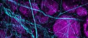 Thinking beyond fluorescence: biological imaging with SHG and THG microscopy
In modern life science research, fluorescence microscopy is a popular and powerful imaging technique. Yet, despite its overall success, the need for fluorescent labels limits its potential applications. How can we image beyond fluorescence microscopy?
However, to improve connections with fellow Italian researchers, I co-founded the Italian Group of Mesenchymal Stem Cells (GISM) in 2014; in 2016 and 2019 I organized the Italian National School of Microscopy and in 2017, I became Italian Ambassador for the European Association for Cancer Research (EACR). Meanwhile, I also became an active science disseminator at events for non-scientific audiences. For example, in 2015, 2016 and 2018 I was the speaker at The Researchers’ Night in Bologna, and in 2015 I was selected as one of the 10 regional finalists of the international FameLab Talking Science Competition, also winning the regional competition in 2017.
Then, wanting to work nearer to centers where data are generated, I decided to move to IRCCS Istituto Romagnolo per lo Studio dei Tumori (IRST) “Dino Amadori” (Meldola, Italy), a cancer institute whose researchers have long-standing experience in anti-cancer in vitro models for drug testing and whose laboratories are equipped with top-notch 3D bioreactors for multicellular spheroids. In other words, a perfect place for extending the experience I had gained in 2D HCS of anti-cancer drugs to 3D models. I am now 36 and have been working as a research fellow at IRST for 5 years, whilst also becoming an Adjunct Professor at the University of Bologna. I have progressed steadily from tutoring a few undergraduate students to being the co-supervisor of two PhD students, also deserving the Italian National Scientific Qualification in FIS/07 – APPLIED PHYSICS and 09/G2 – BIOENGINEERING.
Today, I have 16 conference publications and 38 articles published in scientific journals, including articles in Nature Methods, Nature Reviews Drug Discovery and Nature Communications. I also have two first-author publications in BioTechniques. The fact that I appear as the first or corresponding author in most of my publications (18 first author and eight corresponding author) bears witness to my spirit of independence and leadership qualities to carry out research and achieve my goals. In short, I believe I am a motivated and dedicated researcher and if I won the Future Science Future Star Award, I can promise that I will make excellent use of the provided resources.
What made you choose a career in your field?
At an early age, during my high school studies, I became attracted to electronics and informatics. I had very special professors that led me in developing interesting applications and expanding my knowledge. Already at the age of 19, I was able to create, with LEGO bricks and a microcontroller, a small car that, without any pilot or remote controller, was able to follow a black line on a bright background and transport materials in areas dangerous for humans. The project won several awards and I also published a book on the project: VECPA – Vehicle Electric With Pilot Automatic.
However, I was always attracted to medicine and biology, and at university, I decided to study Biomedical Engineering to connect my passion for electronics and informatics with the life sciences. In particular, for my Master’s thesis, I met Alessandro Bevilacqua, a professor in informatics developing tools and applications for solving technical and biological problems in the field of microscopy. I fell in love with this job and then I decided to spend my life as a researcher in this field!
Describe the main highlights of your career so far
The main highlights of my career can be summarized by listing the freely available tools I designed and share with the community. Here I will report some of those:
2013: MicroMos for building a panorama starting from a set of overlapping images.
2015: CIDRE for correcting the illumination field of microscopy images.
2015: AnaSP software suite to segment brightfield images of multicellular spheroids.
2015: ReViSP for cancer spheroid reconstruction and visualization using a single projection.
2016: CellTracker for tracking 2D cells cultured in vitro.
2017: ReViMS for cancer spheroid reconstruction and visualization using multiple sections.
2017: Advanced Cell Classifier for classifying cells in high-content screening images.
2020: Colour Deconvolution 2 ImageJ/Fiji plugin for stain unmixing in RGB histological images.
2020: 3D-Cell-Annotator MITK plugin for segmenting single cells in 3D datasets.
What is the most difficult challenge you have encountered in your work and how did you overcome it?
I think that defining the ‘most difficult’ challenge is wrong for a researcher: every day I try to overcome research problems. Sometimes the problem arises from a medical doctor needing to analyze patient samples with a microscope, sometimes the problem is highlighted from a biologist observing samples for testing cancer drugs. Honestly, I do not want to define the ‘most difficult’ problem because this would mean to give a rank and all the research problems are difficult and important, and my daily job is to solve all of them, one after the other, hardly working day-by-day, always remembering that “today’s science is tomorrow’s medicine” and so, I’m helping people to have a better life.
Which publication of yours best highlights your career to date?
This publication is the end of a nice story regarding the topic of “illumination correction in microscopy” that started precisely in 2009 when I was 24 years old, with my master thesis, Algorithm for building mosaics of partially overlapping images regarding adherent live stem cells. After 2 years, it brought me to contribute in a conference, then to an article in the Journal of Microscopy; multi-image based method to correct vignetting effect in light microscopy images. In 2013, it led to a second conference contribution: vignetting and photo-bleaching correction in automated fluorescence microscopy from an array of overlapping images. And finally to the publication I mentioned above! Today, the CIDRE tool is known in all microscopy facilities and I’m very proud to have contributed to something that is used worldwide.
What are your main aims for the future?
I want to establish my research group composed of computer scientists and biologists to develop customized tools and applications in the field of microscopy for answering technological and biological questions.
Where do you hope to see yourself in 5 years?
I think that IRCCS IRST is a perfect place for growing and establishing my research group. However, I’m open to other opportunities!
Why do you feel you deserve this award?
I am an extremely motivated researcher, always trying to do my best. The tools I have created are helping a lot of researchers around the world, every day I’m answering emails helping people interested in using these tools. I’m 36 years old and I already have several publications in top-notch journals. I think that I am a good example for other young scientists in the field of biotechniques. Accordingly, I’m happy to be considered for this award!