First-of-its-kind implant could transform tissue loss treatment
Original story from Technion-Israel Institute of Technology (Haifa, Israel).
An implantable tissue flap containing muscle and fat alongside a hierarchical network of blood and lymphatic vessels could offer hope for patients with severe tissue damage.
An international research team led by the Levenberg Laboratory in the Faculty of Biomedical Engineering at the Technion-Israel Institute of Technology (Haifa, Israel) has succeeded in developing a first-of-its-kind, three-dimensional implant that combines muscle and fat tissues, a lymphatic network and a hierarchical blood vessel network. The researchers’ findings were published in Cell Biomaterials.
The current standard treatment for significant tissue loss is harvesting an autologous flap – tissue taken from a healthy area of the patient’s own body – and transplanting it to the damaged site. This approach is used because transplanting tissue from another person leads to immune rejection and associated complications. The new development, therefore, offers an important solution for substantial tissue loss.
According to Shulamit Levenberg, head of the research group, “Our development represents a significant step toward the production of complex implantable tissues for cases involving loss of muscle and fat tissue due to injuries, burns, tumor resection and more. The technology presented in the paper may, in the future, enable the production of personalized flaps tailored to the specific characteristics of an individual patient’s injury.”
A flap is an implant that contains a hierarchical vascular system, a dramatic advantage in terms of integration with the damaged site of implantation. Without such a system, the implant does not immediately receive the oxygen and nutrients it requires, and cellular waste is not properly cleared.
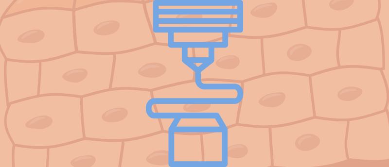
The new flap described in Cell Biomaterials is the first to include a lymphatic network – an essential system that drains fluids from the interstitial space. This flap integrates the lymphatic network together with a blood vessel network and muscle and fat tissues, all within a single structure, along with an arterio-venous loop that connects directly to the blood supply at the implantation site. The complete construct closely mimics the missing natural tissue, including the extracellular matrix, thanks to optimization of the printing process, both in designing the printed structure and in calibrating the syringes that control cell placement within the flap.
From sci-fi to reality: four futuristic advancements in biomaterials science
Explore some exciting preclinical advances in biomaterials science that may, with any luck, prove therapeutically beneficial.
In experiments in rats, the researchers demonstrated that connecting the flap to the target organ led to rapid integration, including continuous delivery of oxygen and nutrients, stable blood flow, normal muscle development and stability of the fat cells. The result: the flap became an integral part of the implantation site and surrounding tissue, both aesthetically and functionally.
The study was led by Levenberg, head of the Technion’s Laboratory for Tissue Engineering and Stem Cells and a world-renowned expert in the development of composite tissue flaps; Eliana Fischer, a physician and graduate of the Ruth and Bruce Rappaport Faculty of Medicine at the Technion and currently a Ph.D. student in the laboratory; and doctoral student Anna Tsukerman, a graduate of the Faculty of Biology who began her Ph.D. after 6 years in the biomedical industry.
The researchers used a unique bio-ink based on components of the extracellular matrix, which mimics the natural tissue environment and enables printing of muscle and fat tissue in a way that promotes cell differentiation. To optimally grow the vascular system, the team printed the engineered blood and prototyped a bioreactor, where they were cultured under flow conditions that mimic natural blood flow in the body and promote maturation of the endothelial layer.
In the next stage, the flaps were implanted in rats and directly connected to an artery and a vein at the target site. The result, as noted, was unprecedented integration of the flap within the body. Notably, although the experiment was conducted in a rat model, the engineered tissues were produced from human cells in order to evaluate the feasibility of the technology for human applications. The group has now begun testing the technology in large animals – the next step toward clinical trials.
This article has been republished from the following materials. Material may have been edited for length and house style. For further information, please contact the cited source. Our press release publishing policy can be accessed here.