Unlocking the potential of graphene in neuroengineering

Original story from the University of California – San Diego (CA, USA).
A safe, non-genetic method for expediting brain organoid growth aids disease research and prosthesis development.
Researchers from University of California San Diego Sanford Stem Cell Institute (CA, USA) have collaborated with scientists at NeurANO Bioscience and Nanotools Bioscience (all CA, USA) to develop and utilize a novel method to stimulate and mature human brain organoids using graphene, a one-atom-thick sheet of carbon. The study introduces Graphene-Mediated Optical Stimulation (GraMOS), a safe, non-genetic, biocompatible, non-damaging way to influence neural activity over days to weeks. The approach accelerates brain organoid development – especially important for modeling age-related conditions like Alzheimer’s disease – and even allows them to control robotic devices in real time.
“This is a game-changer for brain research,” commented co-corresponding author Alysson Muotri. “We can now speed up brain organoid maturation without altering their genetic code, opening doors for disease research, brain–machine interfaces and other systems combining living brain cells with technology.”
A smarter way to grow the brain in a dish
Brain organoids – three-dimensional, stem cell-derived models of the human brain – are valuable for studying neurological diseases, but they usually mature slowly, limiting their usefulness for conditions that develop over decades. Until now, stimulation methods either required genetic modification (optogenetics) or direct electrical currents, which can damage fragile neurons.
GraMOS works by using graphene’s unique optoelectronic properties to convert light into gentle electrical cues that encourage neurons to connect and communicate. This stimulation mimics the environmental input real brains receive, driving development without invasive techniques.
“Using graphene and light, we were able to nudge the neurons to form connections and mature more rapidly, without traditional optogenetic tools,” explained Elena Molokanova, co-corresponding author and chief executive officer and inventor of GraMOS technology at NeurANO Bioscience. “It’s like giving them a gentle push to grow up faster – essential for studying age-related diseases in a dish.”
Key study findings include:
- Faster development: Regular use of GraMOS helped brain organoids form stronger connections, better organized networks and more advanced communication between neurons – even in models made from Alzheimer’s patients.
- Safe and biocompatible: Graphene did not harm neurons or organoid structure, even over long periods.
- Enhanced disease modeling: Early-stage Alzheimer’s organoids revealed functional differences in network connectivity and excitability when stimulated.
- Robotic integration: Graphene-stimulated organoids were linked to a simple robot in a closed feedback loop, enabling it to respond to visual cues.
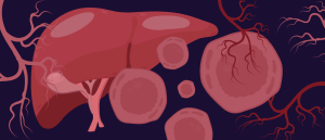 Liver cells self-organize to form vascularized liver organoid
A novel 3D cell culture method has been developed that encourages liver progenitor cells to self-organize and form blood vessel networks for the first time.
From the lab to Alzheimer’s research and beyond
Because stimulation accelerates neural maturation, researchers can study disease progression sooner and in a more physiologically relevant context. This could improve drug testing timelines and provide new insight into how diseases like Alzheimer’s alter brain circuitry.
“Our technology bridges a critical gap in organoid research,” said Alex Savchenko, co-senior author and chief executive officer of Nanotools Bioscience. “It offers a reliable, repeatable way to activate neurons, which can transform both fundamental neuroscience and translational studies.”
Brain meets machine
Brain organoids interfaced with graphene become responsive to their environment and can change their neuronal networks in response to light. This acquired neuroplasticity offers a huge advantage over computer chips in future AI applications by improving the ability of AI systems to solve complex, unforeseen problems and offering greater fault tolerance and reliability in critical applications.
In a striking proof-of-concept, the team connected graphene-interfaced brain organoids to a robotic system equipped with sensors. When the robot detected an obstacle, it sent a signal to stimulate the organoid, which then generated a neural pattern triggering the robot to change course – completing the loop in under 50 milliseconds.
While still far from conscious machines, this integration hints at future neuro-biohybrid systems where living neural tissue and robotics work together for advanced prosthetics, adaptive interfaces or even new forms of computation.
This study is a major step toward unlocking the potential of graphene in neuroscience, nanotechnology and neuroengineering. The technology could lead to new ways of connecting increasingly complex brain-like tissues to each other – and even to the brain itself. The ability to control and accelerate brain organoid development opens the door to using them as powerful models for testing therapies for neurodegenerative and developmental brain disorders, where damaged connections can disrupt the brain’s ability to process and respond to information.
Beyond disease research, the approach could be adapted for tissue engineering, offering a noninvasive, precise way to stimulate other types of lab-grown tissues. And by linking living neural networks to machines, researchers may discover how the brain’s adaptability and learning could enhance computers and robotics – with possible future applications in AI.
“This is only the beginning,” said Muotri. “The combination of graphene’s versatility and brain organoid biology could redefine what’s possible in neuroscience, from understanding the brain to creating entirely new technological paradigms.”
This article has been republished from the following materials. Material may have been edited for length and house style. For further information, please contact the cited source. Our press release publishing policy can be accessed here.