How to print functioning plant cells
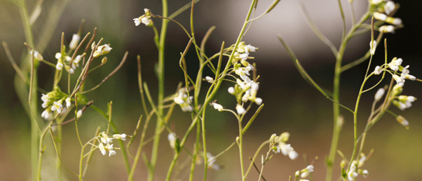
A recent study has established a method to print viable plant cells using a 3D printer.
Researchers at North Carolina State University (NC, USA) have developed a method to 3D print various types of plant cells using bioinks for a reproducible way to study cell-to-cell communication in three dimensions. This method could be used to gain insight into how plant cells communicate amongst themselves and with their environment, which is key to understanding plant cell function. This could eventually lead to improved crop varieties and optimized growing conditions.
“A plant root has a lot of different cell types with specialized functions,” said Lisa Van den Broeck, the first author of the study. “There are also different sets of genes being expressed; some are cell-specific. We wanted to know what happens after you bioprint live cells and place them into an environment that you design: Are they alive and doing what they should be doing?”
Bioprinting plant cells is mechanically similar to 3D printing plastics, but the ink used is made of living plant cells. Additionally, an ultraviolet filter is used to ensure the environment is sterile and multiple print heads are used to print different bioinks at the same time.
Using this technique, the researchers bioprinted live plant cells without cell walls, called protoplasts, from the model plant species Arabidopsis thaliana as well as from soybeans. These were printed with nutrients, growth hormones and agarose. Agarose is a seaweed-based compound that acts as a thickening agent and supports the cells by acting as scaffolding. “We found that it is critical to use proper scaffolding,” explained Ross Sozzani, a co-corresponding author of the paper. “When you print the bioink, you need it to be liquid, but when it comes out, it needs to be solid. Mimicking the natural environment helps keep cellular signals and cues occurring as they would in soil.”
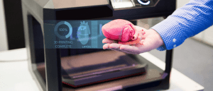 Novel hydrogel bioink improves 3D-printed biomaterials
Researchers are one step closer to bioprinting organs and tissues using a novel granular hydrogel bioink.
Using this bioink, more than half of the printed cells were viable and divided to form microcalli, small colonies of cells. This was the first time that the researchers had maintained cells for longer than a few hours after bioprinting. Van den Broeck added, “similar viability ranges are shown after manually pipetting cells, so the 3D printing process doesn’t seem to do anything harmful to cells.”
This study shows that bioprinting could be used for high-throughput processing. Additionally, this method allows control over the resulting architecture of the cells, for example they could be printed in layers or in a honeycomb arrangement.
The researchers also tested if individually bioprinted cells could divide and multiply. More than 40% of the individual soybean embryonic cells were viable 2 weeks after bioprinting and formed microcalli over time. For optimal viability, the Arabidopsis thaliana root and shoot cells needed different combinations of nutrients and scaffolding. This indicates that 3D bioprinting plant cells could be used to study cellular regeneration in crop plants.
The final part of this study was looking at the cellular identity of bioprinted cells; Arabidopsis thalina root cells and embryonic root cells have a high proliferation rate and can become different cell types, much like stem cells.
“We found that bioprinted cells can take on the identity of stem cells; they divide and grow and express specific genes,” said Van den Broeck. “When you bioprint, you print a whole population of cell types. We were able to examine the genes expressed by individual cells after 3D bioprinting to understand any changes in cell identity.”
Now that a method has been develop to bioprint plant cells that are viable for a long period of time, the researchers plan to study their cellular communication, including at a single-cell level.