Liver cells self-organize to form vascularized liver organoid
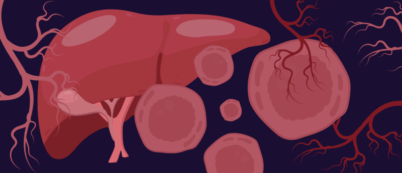
A novel 3D cell culture method has been developed that encourages liver progenitor cells to self-organize and form blood vessel networks for the first time.
The development of accurate liver organoids would enable more in-depth research into the mechanism and treatment of liver diseases. However, the liver has long been a challenge to replicate in organoid form due to its highly specialized vasculature that shapes its metabolic and detoxification functions.
A team of researchers has recently overcome this hurdle, developing a novel 3D culture system that enabled four types of liver progenitor cells to self–assemble into a functional organoid, complete with vascular networks. The research was part of an international collaboration between Science Tokyo (Japan) and Cincinnati Children’s Hospital Medical Center (OH, USA).
 Be picky: selecting cells with desirable characteristics for further development
How do you currently identify and select cells for further growth, characterization and processing? Many available cell-selection technologies lack the sensitivity necessary to preserve cell health and proliferative ability, hindering research workflows and development pipelines. The CellCelector from Sartorius overcomes this limitation, providing an automated system that can identify cells in your culture that possess desirable characteristics and then select and pick them for future development or downstream processing.
Developing the IMALI method
The first obstacle to overcome was the reliable production of liver sinusoidal endothelial progenitors (LSEPs) from human-induced pluripotent stem cells. In this study, the team, led by Takanori Takebe (Cincinnati Children’s Hospital), implemented a careful cultivation protocol and analysis of the key transcriptional changes that occur as stem cells differentiate into endothelial cells.
Once they had a supply of LSEPs, they developed an innovative 3D culture method called inverted multilayered air–liquid interface (IMALI). By arranging multiple cell types in a specialized gel environment, the team was able to induce hepatic endoderm cells, mesenchymal cells, arterial endothelial cells and LSEPs to self-organize into dome-shaped liver organoids.
 Award-winning cancer research: from A to TME
With the rapid progress and high volume of cancer research, awards serve as a good way to keep in mind the foundations of the field and to cut through the noise to highlight the most critical developments in contemporary research. This year’s AACR 2025 (Chicago, IL, USA; 25–30 April), the annual meeting of the American Association for Cancer Research, did just this, to celebrate researchers who have been the driving force behind ground-breaking cancer research.
From mouse to man
Using genetic analysis, the team discovered that these organoids developed four distinct types of blood vessel cells, with sinusoidal vessel cells becoming dominant over time. They also demonstrated that the organoids could accurately replicate some key liver metabolic functions, including producing clotting factors. When transplanted into a hemophilia A mouse model, the organoids and their clotting ability significantly improved bleeding symptoms for up to 5 months.
“This technique provides a foundational method for embedding organ-specific vascular structures into organoids, contributing to the understanding of human biology and disease,” remarked Takebe. “Our enhanced organoids may support the development of regenerative therapies for coagulation disorders and end-stage liver failure, as well as drug discovery and disease modeling.”
The team plans to evaluate the long-term stability and safety of these organoids. They hope that this breakthrough could support advances in personalized medicine and drug discovery.