Map of primate ovarian reserve could enable better disease models
Understanding how the ovarian reserve develops could enable more sophisticated ovarian disease models, aiding treatment development.
Utilizing cutting-edge single-cell sequencing and spatial transcriptomics, a collaborative team of researchers from across the USA has uncovered deeper insights into how the ovarian reserve develops. By improving our understanding of ovary formation, female sex determination and follicle formation, they hope to enable better disease models and revolutionize treatments for infertility and hormonal disorders.
Looking inside the womb
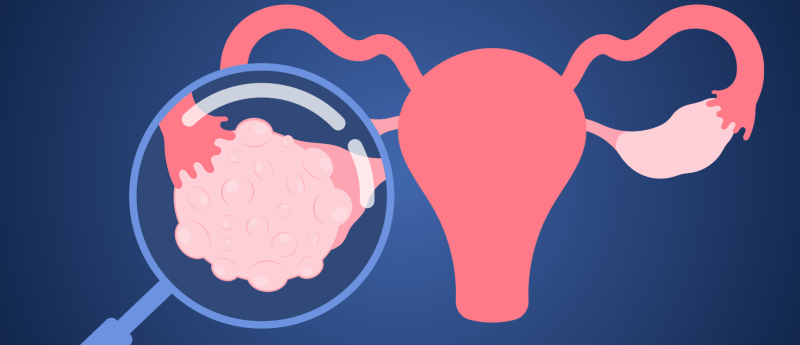
Females are born with a finite egg reserve that is the foundation for reproduction as well as the driver for ovarian hormone production. However, because it is fully formed before birth, scientists have struggled to study it closely.
In a new study, led by Amander Clark from the University of California, Los Angeles (UCLA; CA, USA), a team utilized rhesus macaques as a stand-in for humans. They began by identifying critical stages in ovarian reserve development, then utilized single-cell RNA sequencing to study gene expression at the individual cell level. They applied 10x Visium CytAssist HD and Nanostring CosMx to map gene expression and cell types while preserving the tissue’s spatial organization, as well as microscopy to visualize cellular structures. All data was subjected to rigorous computational analysis to identify key genes and patterns of development.
“Women’s health is already understudied, but the ovary in particular has been neglected,” commented first author Sissy Wamaitha (UCLA). “To effectively treat reproductive health conditions – as well as the growing number of general health issues we now acknowledge affect people with ovaries – we must first develop a fundamental understanding of the full scope of this organ’s function.”
Shining a light on mini puberty
This study has reported the first cellular explanation for why babies undergo a hormone surge shortly after birth, often referred to as ‘mini puberty’. Shortly after birth, the specialist cells that produce hormones activate. The absence of this surge could be the earliest biomarker for polycystic ovary syndrome (PCOS), which affects 10% of women worldwide.
“If we can identify risk factors in infancy that impact ovarian health, then early interventions can be made so that these women don’t suffer once they go through puberty,” explained Clark.
The team hope that these insights will support more accurate ovarian organoids, generating specialized cells from induced pluripotent stem cells that could be combined with lab-grown germ cells to enable future treatment development.