Reference map of retinal pigment epithelium cells created with AI

Researchers identify five distinct cell subpopulations in the retinal pigment epithelium using artificial intelligence (AI), which could explain different severities of retinal degenerative diseases.
A recent study from the National Eye Institute, which is part of the NIH (both MA, USA), analyzed the shape and dimensions of retinal pigment epithelium (RPE) cells using fluorescently labeled images and artificial intelligence. The research team, led by Kapil Bharti, looked at cell morphometry (their size and dimension), hoping to gain insight into the varied spectrum of retinal disease phenotypes.
The RPE is a single layer of cells that sits beneath rod and cone cells at the back of the eye and supports their function. Aging and disease can cause a metabolic change in these RPE cells and lead to photoreceptor degeneration, which impacts vision to varying degrees depending on the location of the RPE cells that are damaged. This includes late-onset retinal degeneration, which affects peripheral vision; and age-related macular degeneration (AMD), where there is damage to the part of the retina responsible for our central vision called the macula. AMD is the leading cause of vision loss.
To investigate retinal disease phenotypes, the team used AI to examine the entirety of the RPE from nine cadaver donors, who had no history of significant eye disease. From this, they were able to produce a reference map of RPE cells, which locates each of the cell subpopulations at a single-cell resolution.
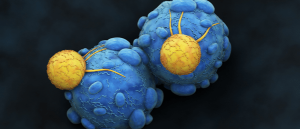 How effective are Killer T cells against COVID-19?
An artificial intelligence-based platform allows researchers to uncover the secrets behind adaptive immunity.
“These results provide a first-of-its-kind framework for understanding different RPE cell subpopulations and their vulnerability to retinal diseases, and for developing targeted therapies to treat them,” said Michael Chiang, who is the director of the National Eye Institute.
On average, 2.8 million cells were analyzed per donor, with 47.6 million analyzed in total. The AI algorithm evaluated the area, hexagonality, aspect ratio (width to height ratio), and the number of neighboring cells. From this, the researchers were able to identify five subpopulations of RPE cells they labeled P1-P5.
These five groups of RPE cells are arranged in concentric circles around the center of the macular called the fovea, which is the most light-sensitive region of the retina. The RPE appeared more tightly packed and perfectly hexagonal around the fovea (P1) than those in the peripheral retina (P4).
The researchers also found that the peripheral retina contained RPE cells with a similar cell area to those around the macula. “The presence of the P4 subpopulation highlights the diversity within the retinal periphery, suggesting that there could be functional differences among RPE that we are currently unaware of,” said Davide Ortolan, the first author of this study. “Future studies are needed to help us understand the role of this subpopulation.”
They also analyzed RPE from cadavers with age-related macular degeneration and noticed a lack of RPE around the foveal, whilst other RPE subpopulations did not have significant differences. Cells that were affected by AMD were observed to be elongated, indicating that different subpopulations are affected by different degenerative diseases. Ortolan commented: “Overall, the results suggest that AI can detect changes of RPE cell morphometry prior to the development of visible apparent degeneration.”
Bharti adds that “the findings will help us develop more precise cell and gene therapies for specific degenerative eye diseases.” These results will inform future studies that use noninvasive imaging technology to examine retinal cells, which could be used to predict changes in RPE health in patients.