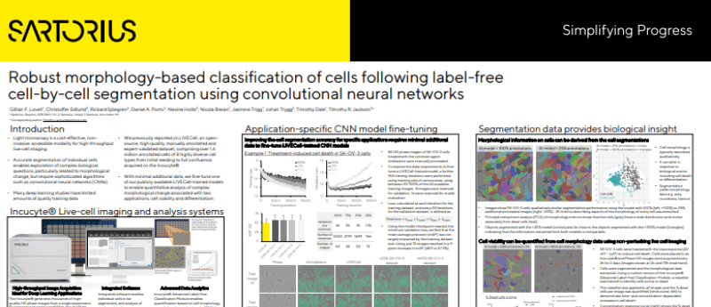
Robust morphology-based classification of cells following label-free cell-by-cell segmentation using convolutional neural networks
Light microscopy is a cost-effective, non invasive, accessible modality for high-throughput live-cell imaging. Accurate segmentation of individual cells enables exploration of complex biological questions, particularly related to morphological change, but require sophisticated algorithms such as convolutional neural networks (CNNs).
Access the poster to see how, with minimal additional data, Sartorius fines-tune one of its publicly available LIVECell-trained models to enable quantitative analysis of complex morphological change associated with two applications, cell viability and differentiation.
Download PosterMore information
In this Poster, you will find:
- Incucyte® Live-cell imaging and analysis systems
- Application-specific CNN model fine-tuning
- Segmentation data providing biological insight
And much more!
This content was provided by Sartorius
You might also be interested in...