Cryo-EM reveals new structural elements in a key ciliary protein complex
Scientists capture the protein complex responsible for transporting signaling molecules to cilia in unprecedented detail.
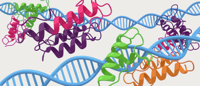
Using single-particle cryogenic electron microscopy (cryo-EM), researchers at St. Jude Children’s Research Hospital (TN, USA) and the University of Texas Southwestern Medical Centre (TX, USA) have solved the 3D structure of a major protein complex in cilia. Their findings serve as a basis for researching developmental diseases involving mutated cilia, which can affect skeletal development, especially the ribs, and structures like the retina.
Cilia are membrane-bound organelles found in nearly all eukaryotic cells and control many signaling processes; however, cilia are unable to synthesize the proteins needed for cell signaling. This means that signaling molecules inside cilia are transported from other parts of the cell. This transport is facilitated by intraflagellar transport complexes called IFT-A and IFT-B. The researchers of this study were able to determine the structure of IFT-A at a resolution of 3–4 Å, visualizing the structure in near-atomic detail.
This revealed details that have not been seen before in IFT-A structures. For example, a previously unknown zinc-binding site, which binds zinc finger protein domains, was revealed in IFT-A. Zinc fingers are critical for certain types of protein–protein interactions; different zinc finger domains are able to interact with DNA, RNA and poly-ADP-ribose, among other proteins. This finding explains some poorly understood connections in the IFT-A complex.
“It was pretty exciting to see the zinc fingers because no one had seen or even predicted zinc-binding sites in IFT-A,” commented co-corresponding author Ji Sun (St. Jude Children’s Hospital). “Our study was able to reveal that with confidence,” adding that this was made possible by the high-resolution structural information obtained in this study.
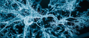 Cilia say what? Important function of neurons’ tiny hairs
Researchers have uncovered a synapse on neurons’ tiny hair-like structures, which may facilitate long-term changes to genomic information in the nucleus.
This study also solved the structure of the IFT-A complex with the Tubby-related protein 3 (TULP3). “TULP3 is a ticket to get on the train of IFT-A,” explained Sun. “TULP3 can then recognize different acceptable cargo to be transported on the train. So, when you have molecular cargo that can hold onto this ticket, it can ride the train. If you disrupt this TULP3 interaction, then you can no longer transport certain cargos, because they lack a valid molecular ticket.” This will give insight into how signaling molecules are transported to and from the cilia, and how disruptions at the interface between IFT-A and TULP3 can cause diseases.
Without knowing the structure of IFT-A, it is challenging to rationalize how changes to any of its six protein subunits can result in disease. This high-resolution structural insight into the IFT-A–TULP3 complex can now be used to study developmental diseases involving cilia and could lead to novel therapeutic approaches to treat them.