Uncovering the cellular origins of cancer and neurodevelopmental disease
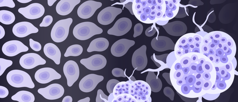
While factors for disease, such as genetic risk, have been extensively identified, their presence alone does not determine progression to disease. To understand what’s driving oncogenesis across diverse populations, multiple factors need to be considered in conjunction: genetics, epigenomics, cell-type-specific pathogenesis and spatial context. A multiomic approach enables the mapping of cellular identity, lineage and interactions within intact tissues, providing a more complete view of development and disease.
 Jasmine Plummer (left) is the founding Director of the Center for Spatial Omics at St. Jude Children’s Research Hospital (TN, USA), where she develops cutting-edge spatial genomics and multiomics technologies to map how cells interact within intact tissues. Her work focuses on uncovering the cellular origins and molecular drivers of cancer and neurodevelopmental disease by integrating high-resolution spatial data with single-cell systems biology.
In this interview, Jasmine shares the techniques she and her lab have developed to investigate the cellular processes underlying disease, some of the limitations of spatial technologies and data analysis tools and the mission of the Global Alliance for Spatial Technologies initiative.
Please tell us about the techniques that you and your lab use to understand the cellular processes that unfold in both normal development and oncogenesis.
The Plummer Lab combines single-cell transcriptomics and epigenomics with cutting-edge imaging-based multiomics, including in situ sequencing, multiplex in situ hybridization and cyclic immunofluorescence, alongside spatial transcriptomics and scalable computational pipelines to map cellular identities and interactions within intact tissues. In collaboration with Luciano Martelotto (then Associate Professor at the University of Adelaide, Australia) and Holger Heyn (Single Cell Genomics Group Leader at the Centro Nacional de Análisis Genómico, Barcelona, Spain), we developed STAMP, a method that transforms standard imaging platforms into single-cell transcriptomic readers by projecting sequencing-based cell identities back onto tissue architecture. Given the size and complexity of spatial omics datasets, we also build advanced computational pipelines for faster, smarter analysis, and have created Spatial Touchstone, a standardization toolkit designed to benchmark and validate samples across platforms in this rapidly evolving field.
Why is studying disease within its spatial context so important? What can spatial approaches tell us that single-cell technologies can’t? How do we get the balance right between capturing spatial context and single-cell data?
Cells function within structured environments, not in isolation. Spatial approaches reveal cell–cell interactions, tissue architecture and microenvironmental niches that single-cell methods alone cannot capture. They show not only which cells are present, but where they are, how they are organized and which neighboring cells or structures may be influencing their behavior. The key is integration: single-cell data defines cell states with high resolution, while spatial data places those states back into their biological context. Importantly, this isn’t about replacing single-cell approaches; those datasets remain incredibly valuable. Many single-cell experiments enable controlled perturbations that are still difficult to achieve with spatial technologies. These perturbation datasets can be used to better inform and train spatial models, improving our ability to interpret spatial patterns and build more predictive, biologically grounded insights.
Once this data is collected, how is it analyzed?
Analysis begins with preprocessing, alignment, segmentation and feature extraction. We then identify cell types or molecular patterns, map them onto tissue structure and analyze spatial relationships such as neighborhoods and cell–cell interactions. The most powerful insights come from integrating spatial data with single-cell, proteomic, imaging and clinical datasets. AI is key to combining these complex modalities, uncovering hidden patterns and building predictive models. Ultimately, this enables faster, more accurate diagnostics by translating multiomic insights into clinically actionable tools for earlier detection and more precise treatment decisions.
 A research revolution: multiomic and spatial techniques in cancer biology
This article explores how the combination of spatial and multiomic methods with machine learning could lead to a deeper understanding of tumor growth and drug mechanisms.
What are the limitations of existing spatial omics technologies and data analysis tools? How do you overcome these limitations?
Current spatial omics technologies still involve trade-offs. Some provide very high molecular depth but lower spatial resolution, while others preserve spatial detail but measure a more limited number of targets. There are also challenges around sensitivity, tissue quality, segmentation accuracy, scalability, cost and cross-platform standardization. On the analysis side, many tools are still evolving, and it can be difficult to compare datasets across technologies or interpret results in a robust, reproducible way. We overcome these limitations through multimodal integration, careful experimental design, strong computational pipelines and collaboration across disciplines. No single platform answers every question, so the most effective strategy is to combine complementary methods and build frameworks that allow results to be validated across datasets and biological systems. Standardization in the field over time will aid in adding robustness to all platforms and data outputs.
Please tell me about the mission of your Global Alliance for Spatial Technologies initiative.
The mission of the Global Alliance for Spatial Technologies is to accelerate the development, adoption and application of spatially resolved technologies across biomedical research. At its core, it is a grass roots initiative that aims to bring together researchers, clinicians, technologists and computational scientists to build shared standards, foster collaboration and lower the barriers to using these powerful tools. It is about creating a global community that can move the field forward more quickly and more equitably, ensuring that spatial technologies are not confined to a few specialist centers but become broadly accessible for answering important biological and clinical questions.
How do you envision advanced spatial omics tools impacting both oncological and neurological research in future?
In oncology, advanced spatial omics will deepen our understanding of tumor heterogeneity, immune organization, metastatic niches and mechanisms of treatment resistance. Paired with AI tools, spatial omics will support more precise biomarkers, better patient stratification, higher accuracy diagnostics and more informed therapeutic decisions. In neurology, spatial tools will be transformative because the nervous system is inherently organized in highly specialized circuits, layers and cellular neighborhoods. These technologies will help us understand how disease unfolds across brain regions, how glial and neuronal interactions change over time and how local microenvironments contribute to neurodegeneration, inflammation or repair. More broadly, I think spatial omics will shift both fields from descriptive cataloging toward a far more integrated view of disease, where molecular changes are interpreted in the context of tissue structure, function and clinical outcome.
The interviewee has no competing interests to report.
The opinions expressed in this interview are those of the interviewee and do not necessarily reflect the views of BioTechniques or Taylor & Francis Group.