Meet the researcher behind Monstrous osteoclasts
 This year, BioTechniques hosted a scientific image competition, and we recently announced that the winning image was Monstrous osteoclasts (see below), submitted by Sayali Chandekar (left). Sayali is a PhD student in the field of biotechnology at the Institute of Bioinformatics and Biotechnology (Savitribai Phule Pune University, Maharashtra, India).
We had the opportunity to follow up with Sayali to hear more about the winning image and her research.
Please can you tell me a bit about your research and how the winning image was captured?
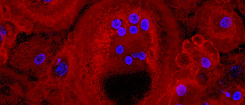
I am studying cellular interactions in the bone marrow microenvironment, under both healthy and diseased conditions. Osteoclasts are bone-degrading cells, which are multinucleated and giant in size. These cells degrade the bone and create space for the formation of new bone. I developed in vitro cultures of osteoclasts from peripheral blood mononuclear cells (PBMNCs) over 21 days. The osteoclasts were then stained for actin by rhodamine phalloidin and the nuclei were stained by DAPI. During one such experiment, the image Monstrous osteoclasts was captured under a Nikon microscope at 20x magnification.
What is the scientific significance of the winning image?
The image was captured from one of the in vitro cultures of PBMNC differentiation. The image gives us an overview of the giant size of osteoclasts and their gross morphology. These osteoclasts are an important cellular component of bone as they play an essential role in bone development. In the process of bone development, the old bone is continuously degraded by osteoclasts and refurbished by osteoblasts (bone-forming cells). As a result, the size and length of bone increases during development along with other physiological processes.
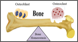
Bone homeostasis maintained by osteoclast (bone-degrading cells) and osteoblast (bone-forming cells).
How does it feel to win this competition?
Contributing to this competition gave me the opportunity both to share my research findings and to get an overview of the research happening around me through the mesmerizing images submitted for judging. Winning a competition always feels great and is a satisfying experience; it feels like I’m getting recognition for all my hard work and research. It gave me a sense of achievement! I also must mention my PhD mentor, Geetanjali Tomar. It’s because of her constant support that my research is progressing.
Monstrous osteoclasts | Sayali Chandekar
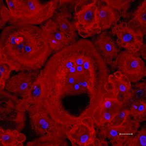
These are bone-eating giant multinucleated osteoclasts generated in vitro from peripheral blood mononuclear cells. The image was taken in 2022 and was taken on a Nikon confocal microscope. The osteoclasts are stained for actin by rhodamine phalloidin and the nuclei were stained by DAPI. The scale bar is 50 μM.
Acknowledgments:
- Central Instrumentation Facility, Savitribai Phule Pune University
- Geetanjali Tomar, PhD Mentor, Savitribai Phule Pune University
- Institute of Bioinformatics and Biotechnology, Savitribai Phule Pune University