Where AFM meets AI: profiling macrophage mechanophenotypes
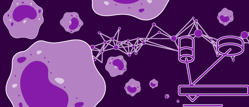
A new method combining AFM with deep learning accurately profiles human macrophage mechanophenotypes.
Macrophages drive key immune processes including inflammation, tissue repair and tumorigenesis via distinct polarization states, the accurate identification of which is vital for diagnosis and immunotherapy. However, methods like RNA sequencing and flow cytometry are often costly, time-consuming and unable to enable real-time, label-free, high-throughput detection.
Atomic force microscopy (AFM) has emerged as a powerful tool in cell phenotyping by decoding the mechanobiological signatures of different cellular states, and AI enables rapid analysis of its complex data. However, macrophages remain underexplored using the combined approach.
In a recent study, a team led by Yang Li from the Shenzhen Institutes of Advanced Technology of the Chinese Academy of Sciences (Shenzhen, China) developed and validated a label-free, non-invasive method combining AFM with deep learning for accurate profiling of human macrophage mechanophenotypes and rapid identification of their polarization states.
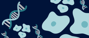 Genomic ‘shake up’ allows cells to play the roles of others
Variability in how DNA is packaged allows cells to take on the identity of different cell types.
Researchers first used localized force-distance curves from AFM to extract biomechanical classifiers, then trained a deep neural network incorporating smart weight assignment and pixel voting to predict macrophage polarization states: naïve (M0), pro-inflammatory (M1) and anti-inflammatory (M2).
To validate the AI model, researchers analyzed the entire population of stimulated macrophages using a weighted voting algorithm originally trained and optimized on well-characterized naïve M0, M1 and M2 phenotypes. The final probability distribution across the four categories (naïve M0, M1, M2 and M1/M2) was 4.3%, 52.2%, 26.1% and 17.4%, respectively.
Moreover, researchers validated the AI model using flow cytometry. They showed that pseudovirus stimulation predominantly induced an M1 phenotype, with smaller proportions of M2 and mixed M1/M2 cells, while naïve M0 cells were nearly absent. The flow cytometry data largely corroborated the classifications generated by the model, underscoring its ability to distinguish macrophage subtypes including mixed phenotypes.
This study provides a powerful tool for probing disease progression and therapeutic responses that can be extended beyond macrophages to other cell types. It paves the way for diagnostics based on mechanophenotypes in cancer, fibrosis and infectious diseases.
This article has been republished from the following materials. Material may have been edited for length and house style. For further information, please contact the cited source. Our press release publishing policy can be accessed here.