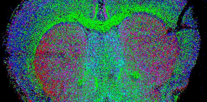
Q&A: spatial single-cell sequencing of tissue samples

CARTANA’s experts answer your questions on in situ sequencing (ISS) following the successful webinar ‘spatial single-cell sequencing of tissue samples.’ Don’t worry if you missed it, you can catch up on demand here.
- Can the in situ sequencing (ISS) method use different tissues?
We have been able to successfully apply our method in different tissues in both humans and mice. This should, however, be determined on a case-by-case basis due to certain tissues requiring specific treatment before sample preparation or sequencing.
- How much optimization should be expected if you apply this technique to tissues other than the brain?
This is highly dependent on the tissue that will be investigated. Certain tissues will require a higher degree of optimization during the sample preparation, in terms of permeabilization for example, and some would require optimization during the sequencing due to increased autofluorescence inherent in the tissue.
- Can one use ISS technology with plant material?
Potentially, yes. However, this would require optimization regarding sample preparation and the actual sequencing. As plant material tends to have a strong autofluorescence signal, this would first need to be overcome before sequencing can be attempted.
- Do you offer probes to survey mesenchymal stem cells in other tissues, such as muscle and bone?
As most of the probe panels are customized to the customer’s specifications, designing probes allowing the survey of mesenchymal stem cells should not pose any issue. It is highly recommended for the customer to already have a candidate list of markers for the probe design.
- Do you have any experience in mapping of glial cells?
We have worked with oligodendrocyte cell types. We also detect general oligodendrocytes, astrocytes and microglia populations, but not subtypes, in many projects.
- Are there any sequencing challenges in glia vs neurons?
Glial cells are more diverse in morphology and physical properties than neurons. Autofluorescence background might be higher in glia. We don’t have enough experience to comment on gene expression levels. However, we do consider most of the foreseeable challenges to be addressable.
- I’m an immunologist, interested in your technique for application to lymphoid tissues sections. Validation of staining patterns revealed by CARTANA would require staining by multiplex immunohistochemistry using panels of antibodies against well-known CD antigens. How would you accomplish this? Have you tried it with brain sections? Has CARTANA been applied to lymphoid sections?
Immunostaining has been shown to be compatible with our current technology in certain cases. This is highly dependent on the application and the stability of the immunomarkers after the ISS procedure. We have had some success with immunostaining in brain sections.
The way immunostaining has been accomplished with our technology is that sample preparation and ISS is performed first at which point the sections will be returned to the customer for immunohistochemistry.
We have not applied our technology to lymphoid tissues but the study done on tuberculosis granuloma [1] might provide some insights.
- Once the slides are stained and imaged, can they then be stained with a morphological stain like H&E and then reimage and overlay the morphological stain with the gene expression data?
Yes, H&E staining can be performed on tissues that have been undergone the whole ISS procedure.
- Can this technology be used to track exogenous cell invasion and integration into tissues following stem cell therapy?
This is certainly an interesting use of our technology. As our probes are highly specific and can detect down to single nucleotide mutations, they should be able to detect exogenous cells and differentiate those integrated into tissues provided they have specific genetic markers that make them distinct from the endogenous cell types.
- You refer to “cell types” — how do you identify specific cells associated with an expression profile? Do you include probes to identify specific cells (neurons, glial, endothelial, etc)? How do you define cell borders to associate a profile with one cell?
We will use ISS to target a combination of genes that define such cell types, followed by comparing the presence/absence and the relative detection level of these genes to the known expression profile.
We often include detection of the general types like the ones mentioned here. Compared to detecting cell subtypes, this is much easier to achieve and often requires 2-3 broad markers per type.
Currently, we define cell borders based on nuclear segmentation and expansion. Therefore, targeted transcripts should preferably be residing in cell soma.
- How will this technique help in screening the cancer cell? For cells that have barcoding, we can easily go for probing. What can be done in state-of cells which is unique to the mutation?
Our probes are highly specific and can be used to detect mutations at a single nucleotide resolution. There are a couple of papers that describe cancer mutation detection using our technology [2,3].
- What about the previous processing of the sample? Is it necessary to have frozen intact tissue? Do you need to perfuse the animal before? Do you need frozen and FFPE sections?
Both types of tissues can be used with our technology. Our experience is that fresh frozen sections yield much better results, meaning higher sensitivity.
- What is the range of thicknesses of tissue that have been tried? Does the method work with standard ~5-micron FFPE sections?
Usually, sections with a 10-micron thickness are used for our technology. A range between 5-25 microns are feasible to sequence. Higher thickness can pose challenges for the distribution of signals over the thickness of the tissue.
- Have you tried to detect viral RNA using the ISS method?
There are indeed a couple of studies that describe the detection of viral RNA using our technology. We can refer you to a paper that uses our library preparation scheme to label Influenza infections [4]. The use of ISS will allow higher multiplexing, which could include some immune host response markers.
- You indicate low false positive rate -what about false negatives given you do not do amplification which could result in false negatives for low copy number genes?
There is an amplification step included in our sample preparation scheme. However, since our technology relies on a reverse transcription step the sensitivity towards low expressed genes is currently one of our limitations. A way to overcome this is to use a larger number of probes for those genes.
- Is the wet lab work manual or could it be performed on an automated staining platform similar to RNAscope?
At the moment the wet lab work is manual, however, some slide staining robots can be used to automate the sample preparation steps. We are currently working together with another company to build an automated solution for the sequencing part of our protocol.
- How much does it cost?
Information about price can be received sending a request on our website form.
Don’t miss out – catch up on ‘spatial single cell sequencing of tissue samples’ and learn more about ISS technology by registering for the webinar on demand now.
About CARTANA
CARTANA is a Swedish spatial genomics company that has been spun-out from Mats Nilsson’s lab at Science for Life Laboratory (Stockholm, Sweden) that commercializes the ISS method, which has been developed in the Nilsson lab over decades. ISS quantitatively measures expression of hundreds of genes in a single tissue section at single-cell resolution. CARTANA offers both kits and ISS service.
For further information, please contact Francesca Bignami, Chief Marketing Officer.