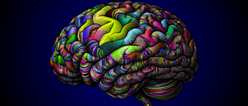
A micron-resolution map for navigating tissue fiber networks
A new method has revolutionized our ability to map tissue fiber orientation and organization across tissues, diseases and sample preparations.
An international group of researchers led by Marios Georgiadis from Stanford Medicine (CA, USA) has recently developed a simple, low-cost method for mapping tissue fibers at micrometer resolution. Originally designed to analyze brain sections, the novel method – called computational scattered light imaging (ComSLI) – has provided insights into neurodegeneration and demonstrated potential for analyzing tissues beyond the brain, contributing to a broader understanding of health and disease.
Tissues are composed of networks of microscopic fibers, each with a specific orientation and organization that contributes to the tissue’s specialized function: for example, muscle fibers coordinate mechanical forces and brain fibers transmit signals that underlie cognition. However, existing methods for visualizing these fibers – such as diffusion MRI, electron microscopy and small-angle X-ray scattering – have drawbacks, forcing researchers to compromise when it comes to resolution, tissue volume, expense or sample preparation.
To overcome the limitations of current methods, the research team developed ComSLI, an optical microscopy method that utilizes a directed rotating LED light and a high-resolution camera to recover fiber orientation maps from animal and human tissues, regardless of their age, storage conditions or the staining protocol used. ComSLI works under the principle that light scatters differently depending on the orientation of the material it passes through. By rotating this LED light and recording how that affects scattering, software algorithms can then recognize the scattered light patterns and produce color-coded maps that indicate the density and orientation of fibers within a sample.
This method is particularly celebrated for its simplicity and flexibility. “This is a tool that any lab can use. You don’t need specialized preparation or expensive equipment. What excites me most is that this approach opens the door for anyone, from small research labs to pathology labs, to uncover new insights from slides they already have,” commented co-senior author Michael Zeineh (Stanford).
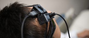
Speckle contrast optical spectroscopy: a solution to stroke screens?
A multidisciplinary team of researchers has developed a device dependent on speckle contrast optical spectroscopy to rapidly measure brain blood flow.
Applying ComSLI to their study of neurodegeneration in the hippocampus, the team compared entire formalin-fixed, paraffin-embedded brain tissue samples from a patient with Alzheimer’s disease and a healthy individual. They found that the Alzheimer’s sample displayed significant deterioration in the hippocampus, with the perforant pathway – a key memory-related signaling pathway – almost undetectable, which presented a stark contrast to the densely fibered healthy brain sample.
To test the new method’s versatility for analyzing tissue samples of different ages and varied preparations, the team used ComSLI to image a brain section prepared in 1904. The technique successfully captured intricate fiber pathways, demonstrating its potential for studying historical samples and tracing disease lineages.
Although developed for neuroimaging, ComSLI is also applicable to other tissues, including bone, muscle and vascular samples. This ability to map intricate fiber orientation and organization across tissues, diseases and sample preparations provides researchers with an invaluable tool for deepening our understanding of pathology as well as the relationship between tissue structure and function.
“Although we just presented the method, there are already multiple requests for scanning samples and replicating the ComSLI setup – so many labs and clinics would like to have micron-resolution fiber orientation and micro-connectivity on their histology sections,” Georgiadis concluded. “Another exciting plan is to go back to well-characterized brain archives or brain sections of famous people, and recover this micro-connectivity information, revealing ‘secrets’ that have been considered long lost. This is the beauty of ComSLI.”