Achieving novel 3D imaging of lipids with Lipid-CLEM
An international research team presents a new imaging technique to make lipids in cellular membranes visible and show how they are organized at the nanoscale.
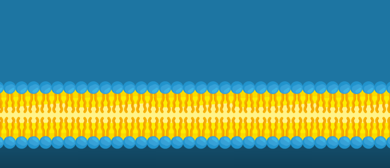
Biological membranes of cells and its subunits (organelles) are organized into tiny regions (nanodomains) made up of fats (lipids) and proteins. Those specialized regions carry out important tasks for the cell, such as signaling, sorting or transport. While proteins in these domains are well understood, the lipid distribution and behavior within them remain a bit of a mystery, as lipids move very quickly and existing methods struggle to visualize individual lipid species at high resolution.
To localize lipids, researchers use ‘bifunctional lipid probes’, which are very small, slightly modified lipids that act like molecular GPS tags. These probes can be added into living cells, then ‘frozen in place’ with light (photo-crosslinking), and later labeled with fluorescence using a chemical reaction (click chemistry). In this way, researchers can track where specific lipids are and not alter and disturb the cell too much.
However, light microscopy alone is not enough to visualize small details in the cell membrane. Higher details can be captured by electron microscopy. Correlative light and electron microscopy (CLEM) combines the strengths of both techniques. Together with the bifunctional lipid probes, Lipid-CLEM shows where labeled lipids are and makes the fine structure of the membranes visible.
Previous CLEM methods, though, either damaged the membrane structure, only worked on the outer surface of the cell or couldn’t distinguish individual lipid species. To fix these issues, Mathilda Lennartz and a team of researchers in the group of André Nadler, group leader at the Max Planck Institute of Molecular Cell Biology and Genetics (MPI-CBG; Dresden, Germany), and the group of Ori Avinoam at the Weizmann Institute of Science (Rehovot, Israel) have now developed a new method that they call Lipid-CLEM.
 Put a ring on it: redesigning the delivery vehicles behind mRNA vaccines
The modification of lipid nanoparticles with aromatic rings and disulfide bonds enhances mRNA vaccine delivery.
Novel 3D imaging of lipids
Mathilda Lennartz, co-corresponding and lead author of the study, explained, “To study lipid sorting in early endosomes – a key sorting station inside the cell – cells must be rapidly frozen to stop lipids in their tracks and to preserve the membrane of the cells. Later, these lipids can be labeled on very thin slices of the sample, termed ‘sections’, of cells using click chemistry. These sections are what we then image using the Lipid-CLEM approach.”
“With Lipid-CLEM, we observed that a specific lipid called sphingomyelin is more common in small vesicles inside the endosome and less common in tubular membrane domains. This separation has already been observed for some proteins,” shared Mathilda. “What we concluded from this is that at least some lipids, just like proteins, must also be sorted in the endosome. Interestingly, in our study, sphingomyelin and a protein cargo arrive at the same time in the early endosome but separate into different domains, indicating that lipid and protein trafficking routes can diverge during this sorting.”
The power of team work
Ori Avinoam’s team at the Weizmann Institute brought in their expertise in correlative light and electron microscopy to this study. Ori commented, “This study highlights how essential collaborations are for driving research forward. Bringing together complementary expertise allowed us to establish a method that made it possible to uncover fundamental principles of lipid sorting that were previously inaccessible.”
André Nadler, corresponding author, concluded, “Our Lipid-CLEM workflow enables 3D visualization of lipid densities in membrane nanodomains, offering a new way to study lipid organization in complex cellular structures. We finally can look at lipid sorting in membranes with the resolution we need. We believe that our new method Lipid-CLEM will help us to better understand how lipids work in cells, as it allows us to study both lipids and proteins together during membrane organization and function. This may also contribute to a better understanding of membrane dysfunction-related diseases.”
This article has been republished from the following materials. Material may have been edited for length and house style. For further information, please contact the cited source. Our press release publishing policy can be accessed here.