Infographic: Investigating biomechanics with atomic force microscopy
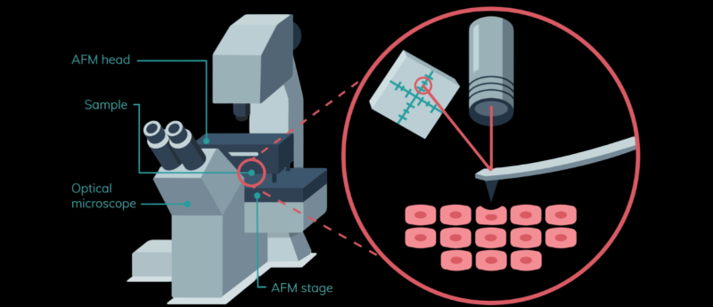
In this infographic, we dive into how atomic force microscopy (AFM) works, the technical features one must consider and how it can be implemented for biomechanical investigation.
AFM provides high-speed structural imaging and nanomechanical measurements to enable crucial insights into the relationship between structure, morphology and function at the cellular and molecular level, both in normal and diseased states.
This infographic is part of the BioTechniques In Focus on AFM, in association with Bruker BioAFM. Check out the animated video to learn more.
This feature was supported by Bruker BioAFM.
![]()
You might also be interested in...