How does cholera colonize the gut? Unmasking virulence activation with cryo-EM
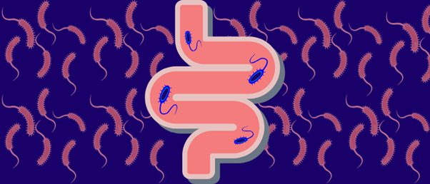
Cryo-electron microscopy (cryo-EM) has heralded a structural biology breakthrough that advances our understanding of how Vibrio cholerae colonizes the human gut and causes often life-threatening disease.
In an international collaboration, researchers from the Institute for Research in Biomedicine, the Institut de Biologia Molecular de Barcelona (both Barcelona, Spain), European Molecular Biology Laboratory Heidelberg (Germany) and the University of Detroit Mercy (MI, USA) have used cryo-EM to resolve several structures within the transcription activation complexes of Vibrio cholerae – the bacterium behind cholera. In doing so, they have unveiled the molecular mechanism underpinning cholera virulence gene activation, which could be exploited to design novel drugs to treat the disease.
Cholera remains a major global public health threat, with between 1.3 and 4 million cases estimated worldwide each year. To effectively combat the disease, we need to understand how the pathogen responsible for it behaves inside the body.
The V. cholerae virulence activation cascade has been thoroughly studied. For example, we know that the transcription factors ToxR and TcpP bind to the ompU and toxT promoters, ultimately resulting in the expression of the cholera toxin and the toxin coregulated pilus – a colonization factor that anchors the bacteria to the intestinal walls. However, little is known about how RNA polymerase (RNAP) is recruited to kick off the process, the interactions between RNAP and these transcription factors or the differences in activation mode of each promoter.
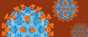 Antiviral insights: cryo-EM and optical tweezers reveal more about drug–herpes virus interaction
Cryo-EM images show drugs bound to herpes simplex virus protein at nearly atomic detail, while optical tweezer experiments show how the drug-bound protein behaves in real time.
In an attempt to change this, the team used single-particle cryo-EM to solve five structures in the ompU and toxT transcription activation complexes, including the RNAP holoenzyme, promoter DNA and their corresponding transcription factors, ToxR or TcpP and ToxR-TcpP, respectively.
The visualization revealed that virulence activation is achieved through the interaction of ToxR or TcpP with the α–C-terminal repeat domain of RNAP, where a single amino acid of the activator, a phenylalanine, appears to act as a critical molecular bridge. This was later confirmed by mutagenesis.
Both ToxR and TcpP transcription factors display similar, but not identical, activation mechanisms. Small differences in their structures, interaction and fine-tuning mechanisms result in different promoter selection for the ompU promoter, and a collaborative activation for the toxT promoter.
The researchers already knew that the structures of the components of the holoenzyme were very similar to those of the Escherichia coli RNAP, as the sequence is highly conserved between both species of bacteria. However, unlike in E. coli, the binding of the transcription factor doesn’t cause a component rearrangement of the polymerase. Instead, it stabilizes the RNAP–DNA interaction.
The cryo-EM structures, together with the team’s previous work on the regions immediately upstream of both promoters, suggest a model that explains the virulence-related transcriptional cascade activation in V. cholerae, after the bacteria reach the intestine.
“Understanding this interaction at the molecular level gives us a new way to think about how bacterial virulence is controlled,” commented lead author Miquel Coll (The Institut de Biologia Molecular de Barcelona).
Furthermore, this could open up the possibility of new therapeutics. The similarities between E. coli and V. cholerae RNAP may mean that antibiotics proven effective against the former, such as rifamycins, can also target the latter, and potentially even tackle multidrug resistance in V. cholerae.