Meet the researcher behind the 2025 winning image
Our annual scientific Image Competition strives to showcase the beauty and intricacy of the life sciences through images captured in the lab and in the field. We had several exceptional submissions this year, ranging from pictures of zoanthids to fluorescent Klebsiella pneumoniae. This year, the winning image was Human neurons reprogrammed from skin cells, submitted by Bruno Cisterna.
Bruno Cisterna is a neuroscientist and microscopist in the lab of Eric Vitriol at Augusta University (GA, USA). His research focuses on how the cytoskeleton contributes to neurodegenerative diseases such as amyotrophic lateral sclerosis and Alzheimer’s disease, using cell culture models derived from induced pluripotent stem cells. He is especially interested in how changes in microtubules and the actin cytoskeleton affect the health and communication of neurons. Along with his scientific work, Bruno also explores the artistic side of microscopy, capturing images that reveal the hidden beauty of cells.
In this interview, Bruno shares more about his winning image, his favorite microscopy techniques and his advice for burgeoning microscopists.
Please tell us about the winning image, Human neurons reprogrammed from skin cells.
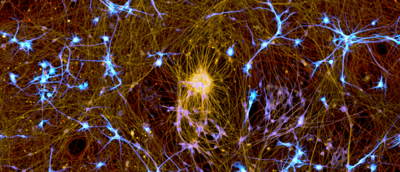
This image shows a cluster of human neurons that were reprogrammed from skin cells and matured for 24 days. The cyan color highlights microtubule-associated protein 2, the yellow shows microtubules and the red marks acetylated microtubules. I captured it using a Nikon CSU-W1 SoRa spinning disk confocal microscope with a Hamamatsu ORCA-Fusion BT camera at 20x magnification.
What stood out to me while working with this stem cell-derived neuron model is how much coordination the cells begin to develop as they mature. Even though their neurites extend in many directions, they still maintain regular patterns in how they organize themselves. For example, dendrites often space out in a surprisingly even way. It feels like a kind of order within the apparent chaos of neuronal projections, and it is not something you usually observe in immortalized cell lines. Watching that organization emerge over time has been one of the most fascinating parts of this work.
Which microscopy technique is your favorite and why?
Throughout my scientific career, I have had the opportunity to explore many microscopy techniques, and each one has its own strengths. Lately, I have been working a lot with spinning disk confocal microscopy, especially the SoRa system, which offers super-resolution capabilities. It allows me to capture delicate neuronal structures in great detail while still being gentle enough for sensitive samples. I have also become very interested in refractive index-based microscopy because of its simplicity and versatility, particularly for exploring different types of samples and structures.
What interests you most about microscopy?
What I enjoy most about microscopy is how visual it is. It helps make complex biological processes feel more real and easier to connect with. I love the moment when something hidden becomes visible for the first time. Being able to share those images with others, so they can see that beauty too, is a big part of what makes it fun for me.
What’s one piece of advice you’d give to someone wanting to better their microscopy skills?
My main advice is to slow down and really spend time with your sample. Try different settings, play with the light and look closely at the fine details. Good images often come from patience and curiosity. The more time you spend observing, the more you start to see and the more rewarding microscopy becomes.
The interviewee has no competing interests to report.
The opinions expressed in this interview are those of the interviewee and do not necessarily reflect the views of BioTechniques or Taylor & Francis Group.