Visualizing the invisible: 5 recent breakthroughs in microscopy
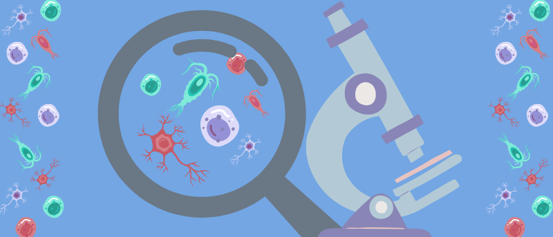
Microscopy is an imaging technique that enables us to see a world that would otherwise be invisible to us.
Once upon a time, visualizing cells, microbes and other entities not perceptible to the naked eye was impossible. Then came the advent of microscopy in the 16th century, which brought the minuscule world around us into sharper focus. Microscopy has come a long way in the centuries since, transforming our knowledge of life on the microscopic scale and heralding breakthroughs across the life sciences.
Here, we explore five recent applications and innovations in microscopy that have magnified our understanding in the fields of microbiology, neuroscience and cell biology.
Super-resolution sectioning image scanning microscopy: visualizing biological tissues in unprecedented detail
Super-resolution sectioning image scanning microscopy – a new optical microscopy technique devised by scientists at the Istituto Italiano di Tecnologia (Genoa, Italy) – has enabled researchers to visualize and photograph dense, complex biological samples in extremely sharp detail.
This is something that has proven difficult in the past. Using confocal microscopy to image such samples means a trade-off between spatial resolution and signal-to-noise ratio. Image scanning microscopy overcomes this, but current approaches do not provide optical sectioning and often fail with thick samples.
That’s where super-resolution sectioning image scanning microscopy comes in. The novel technique has improved spatial resolution and contrast when studying thick tissues. Sensors capture light when it hits the sample and detect variations as the light spreads throughout it. Then, a specially designed reconstruction algorithm inverts the physical model of image formation.
The result is an image with digital and optical super-resolution, high signal-to-noise ratio and enhanced optical sectioning.
“The optical microscope used is equipped with an array of SPAD detectors (single-photon avalanche diode), capable of detecting the arrival of individual photons with very high spatial and temporal precision,” explained Alessandro Zunino, first author of the study.
“This characteristic not only improves the resolution and optical sectioning, but also enables advanced techniques such as fluorescence lifetime, which are fundamental to explore molecular dynamics in living tissues and to provide functional as well as structural information.”
The new technique has been made available to the scientific community following the principles of open science. Potential applications are numerous, from studying disease to investigating how drugs interact with biological tissues.
Read more here | Press release
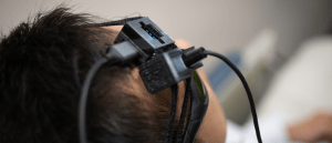
Speckle contrast optical spectroscopy: a solution to stroke screens?
A multidisciplinary team of researchers has developed a device dependent on speckle contrast optical spectroscopy to rapidly measure brain blood flow.
Ultrastructure expansion microscopy: revealing the inner workings of invisible life
A team led by researchers from the European Molecular Biology Laboratory (Heidelberg, Germany) and the University of Geneva (Switzerland) has used ultrastructure expansion microscopy (U-ExM) to peek at the cellular architecture of hundreds of marine plankton species.
Visualizing the inner structures of such microscopic organisms has always been a challenge for biologists. Conventional light microscopes lack the necessary resolution, while transmission electron microscopy and, more recently, volume electron microscopy, are limited by throughput, cost and labeling constraints.
However, expansion microscopy, later refined into U-ExM, offers a solution. Using water to physically enlarge biological samples and make an organism’s cell wall permeable, the technique enables the visualization of preserved ultrastructures by optical microscopy.
In the latest study, the team used U-ExM to carry out high-resolution volumetric imaging of over 200 planktonic eukaryotes. When combined with pan- and specific immuno-labeling, they were able to map microtubule and centrin organization and assign molecular identities to cytoskeletal structures previously only observed by electron microscopy.
Their findings demonstrate how expansion microscopy can be instrumental in yielding biological insights into evolution and cellular function.
“U-ExM is transforming how we explore protist ultrastructure,” remarked Armando Rubio Ramos (University of Geneva), co-first author of the study. “By combining this technique with high-throughput imaging and comparative analyses, we can begin to decode how cellular architecture has diversified across evolution. It’s a bridge between molecular data and the physical organisation of life at the microscopic scale.”
Read more here | Press release
 A micron-resolution map for navigating tissue fiber networks
A new method has revolutionized our ability to map tissue fiber orientation and organization across tissues, diseases and sample preparations.
Miniaturized widefield microscopy: imaging real-time neuron activity in awake animals
A new miniature widefield microscope that can capture real-time voltage signals in free-moving rodents has been designed by researchers at the University of Colorado Boulder (CO, USA), University of Colorado Anschutz Medical Campus (CO, USA) and Columbia University (NY, USA).
Genetically encoded voltage indicators (GEVIs) – fluorescent proteins that can be used to optically track electrical activity – are an exciting tool in neuroscience. They can be used to observe voltage spikes as an indicator of neural computation in awake animals, and as such, give scientists insight into how brain cells process information during natural behavior. However, voltage imaging in freely moving animals faces various technical limitations, hence the need for novel technologies that facilitate GEVIs to capture the moment a neuron fires.
Miniature microscopes are one approach researchers are using to tackle this. In this recent development, the team designed MiniVolt – a compact widefield microscope designed for voltage imaging. The custom-designed optical system has a numerical aperture of 0.6, a 250 µm field of view, a 1.3–1.6 mm working distance and weighs just 16.4 g. It can image in vivo voltage spikes at a rate of 530 frames per second with a spike peak-to-noise ratio greater than 3.
In experiments with mice, they demonstrated that the tech could image real-time neuron activity in awake animals, and that the voltage recordings closely matched those from a standard widefield microscope.
“By capturing these detailed voltage patterns across different parts of the brain, our microscope makes it possible to directly explore how subtle electrical signals influence the timing of brain activity, such as spatial navigation in the hippocampus,” lead author Emily Gibson (University of Colorado Anschutz Medical Campus) commented. “An increased understanding of how neural circuits guide behavior and cognition could lead to new treatments for a variety of neurological disorders and neurodegenerative diseases.”
Read more here | Press release

How do flu viruses infect cells? New microscopy method captures complex ‘dance’
How flu viruses enter cells has been directly observed thanks to a new microscopy technique with the potential to revolutionize research on membrane biology, virus–host interactions and drug discovery.
Curvature-adaptive gigapixel microscopy: capturing non-flat samples
Scientists at Duke University and Ramona Optics Inc. (both NC, USA) have designed a new microscope that captures large, high-resolution images of curved samples in a single snapshot.
Traditional microscopes assume that a sample is perfectly flat, but in real life, this is rarely the case. A common workaround is to use mechanical scanning to ensure focused capture at reduced throughput, which slows down the imaging process.
In an attempt to remedy this, the researchers created PANORAMA: a single-shot, re-imaging multiple-camera system that acts like a single giant microscope. It combines a telecentric lens with a large-aperture tube lens that projects an image of the sample onto a micro-camera array, which images different portions of the sample. The images from each camera are then stitched together into a continuous picture.
As a result, PANORAMA achieves seamless, gigapixel imaging and submicron resolution even if the sample is curved, eliminating the need for scanning.
“With our approach, it’s possible to adjust the focus across the sample, so that everything remains in focus even if the sample surface isn’t flat, while avoiding slow scanning or expensive special lenses,” explained team leader Roarke Horstmeyer (Duke University).
The microscope could be used in medical pathology to scan tissue slides, as well as in materials science or industrial inspection to inspect large surfaces.
Read more here | Press release
 Probing the brain for answers: a PET tracer for early Alzheimer’s diagnosis
In this interview, we learn about how Yanyan Zhao and her colleagues have approached the preclinical development of a PET imaging tracer for catching Alzheimer’s disease earlier.
Time-deterministic cryo-optical microscopy: snapping frozen cells
An international team led by Osaka University (Japan) has developed time-deterministic cryo-optical microscopy – a technique to freeze biological cells in milliseconds during optical microscopy observation.
Achieving high-resolution, accurate snapshots of dynamic cellular activity is no mean feat. Fluorescence microscopy techniques are widely used in this area of research, which requires rapid imaging to capture constantly changing processes. Unfortunately, attaining a high signal-to-noise ratio under such short image acquisition times remains a challenge.
Time-deterministic cryo-optical microscopy combines the advantages of live-cell and cryofixation microscopy – for example, temporal dynamics and high signal-to-noise ratio snapshots – to rapidly freeze samples while more effectively preserving cell morphology and condition.
To analyze the data obtained with this new microscopy method, study author Rainer Heintzmann provided a 3D reconstruction software that transforms raw data into detailed 3D images, revealing cellular structures and molecular processes with incredible clarity.
The combination “opens fascinating insights into cellular processes that were previously blurred or invisible,” Heintzmann concluded.