Viewing macrophages with a novel ultrasound imaging technique
Using a bubble-based technique, researchers can now image macrophages in situ.
Researchers at Penn State University (PA, USA) have developed an ultrasound imaging technique for continuously viewing macrophages in mammalian tissue. Macrophages bridge the gap between innate immunity and adaptive immunity, regulating both types of responses and playing a key role in human health. Ultrasound is often used for visualizing internal tissues; however, this imaging technique can’t distinguish macrophages from other cells. By developing a way to visualize macrophages and gain a better understanding of their behavior in the body, researchers hope to harness these cells for therapeutic use.
Macrophages are immune cells that are involved in pathogen detection and clearing, wound healing and inflammation mediation. Due to their significant involvement in regulating our immune responses, they could be instrumental players in developing therapies for numerous conditions, such as cancer, infections and autoimmune disorders. Before we can start developing these therapeutics, however, we need to be able to visualize them properly.
“If we could visualize what these cells are doing in the body, in real-time, then we could learn a lot about just how diseases progress and how healing happens,” senior author Scott Medina commented. “This would give us a view of what the cells are doing in the body because right now, we’re really limited to taking the cells out of the body and seeing what they do in a petri dish, which is not going to be the same behavior that we see in the body.”
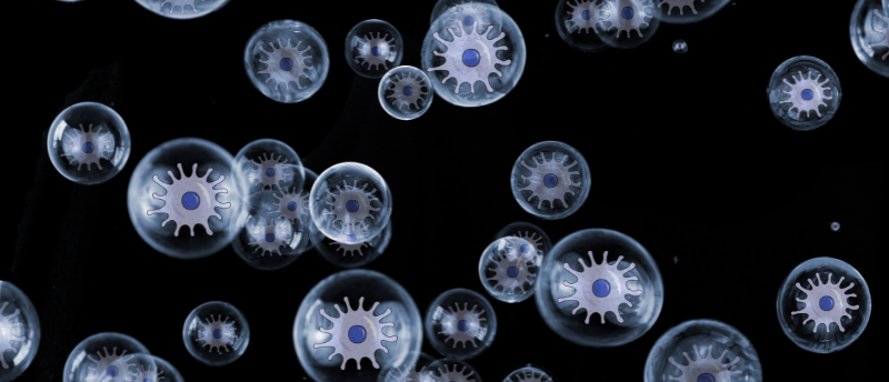
 Get in formation: ultrasound waves direct cells into place
Researchers have developed an alternative to optical tweezers using ultrasound to push and pull cells selectively, advancing cell sorting abilities.
To visualize macrophages using ultrasound technology, the team needed to create a contrast agent, a label that could tag the macrophages and make them stand out from the rest of the cells. To do this, they developed peptide nanoemulsions that undergo a phase change during ultrasound imaging. The phase change from nanoemulsion droplets to gas bubbles occurs due to the change in pressure caused by the ultrasound waves. The gas bubbles then reflect the ultrasound waves, providing a contrast between the labeled macrophages and the surrounding cells.
When testing this technique in porcine tissue, the researchers found that this novel ultrasound imaging technique effectively distinguished macrophages from the other cells in the tissue.
This technique allows researchers to observe how immune cells act in the body, tracing their movements and activities. By understanding how the immune system is regulated and how it fights disease, the team hopes to develop better immunotherapies in the future. Additionally, the team wants to collaborate with others to develop the technique further to visualize other types of immune cells or monitor plaque buildup in arteries.