Who won the 2025 Nobel Prize for Physiology or Medicine?

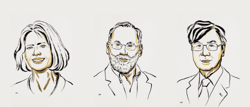
The discovery and characterization of regulatory T cells has been selected as a Nobel Prize winning research, with three researchers selected to share the prize.
Mary Brunkow (Institute for Systems Biology, WA, USA), Fred Ramsdell (Sonoma Biotherapeutics, CA, USA) and Shimon Sakaguchi (Osaka University, Osaka, Japan) have been awarded the Nobel Prize in Physiology or Medicine 2025 for their discoveries concerning regulatory T cells (Tregs) and the genetic factors that underlie peripheral immune tolerance, which prevents the immune system from harming the body.
Going against the flow
In the ever-evolving arms race between pathogens and the immune system, some pathogens have evolved to present molecular structures that mimic those found in the host, and while the receptors in host immune systems have evolved to detect them, the sheer number of receptor/substrate combinations mean that inevitably some of these receptors will misfire and bind to healthy somatic cells rather than pathogenic ones. So, in this chaotic warzone, how does the immune system avoid spiraling rapidly out of control?
For many decades, the answer to this question was solely attributed to the process of central tolerance. During this process, recently produced T cells, transported to the thymus for maturation, are screened past medullary thymic epithelial cells (mTECs) that present tissue-specific antigens. Any T cells that have produced receptors capable of binding to the host’s own antigens are caught by the mTECs and undergo apoptosis.

Illustration of the process of central tolerance in the thymus by Mattias Karlén. © The Nobel Committee for Physiology or Medicine.
This neat, ELISA-like process proved satisfactory for most researchers, and early investigations into potential ‘suppressor T cells’ in the 70s were limited by the technologies available and were constantly dogged by methodological inconsistencies and contradictions in the research. The field suffered what could have been a fatal blow when, in 1983, new sequencing technologies disproved the existence of the ‘I-J locus’ in mice, which was theorized to be a lynchpin of these suppressor T cells.
Despite the dismissal of this field by the wider community, Sakaguchi continued to investigate, convinced that there was more to the story. Previous work in the 70s had shown that mice that had their thymus excised would develop autoimmune thyroiditis, as expected, but that by injecting them with lymphocytes from healthy mice, this condition could be prevented. This indicated that a cell population in this infusion was acting to dampen the autoimmune effects of rogue T cells.
Sakaguchi, working in 1982, repeated and remoulded these experiments, this time using a subset of cells that expressed the surface markers CD5 and CD45RB, drawing the same conclusions. This work garnered new attention for the field, and with the advent of monoclonal antibodies for cell surface antigens enabling improved characterization of cells, the hunt for a suppressor T cell was reinvigorated.
Building on previous studies that showed CD4+ T cells with low levels of CD45RB had a protective effect against autoimmune conditions, Sakaguchi continued to further characterize and subdivide these cells, taking into account a wider selection of surface markers. This came to fruition in his 1995 study, in which he took two cohorts of athymic mice and administered one with a population of CD4+ cells that lacked any cells that also expressed CD25+, and the other with a mixed population of CD4+ and CD4+CD25+ cells. The CD25+ depleted cohort became sick, while the cohort that received the CD4+CD25+ cells remained healthy. Sakaguchi had identified his cell and the term ‘regulatory T cell’ took off.

Illustration of the experiment highlighting the role of CD4+CD25+ T-cells in rescuing athymic mice by Mattias Karlén. © The Nobel Committee for Physiology or Medicine.
CD25 was, however, also expressed at lower levels in some effector T cells, making it a limited marker for these cells, and some doubt still remained. The hunt had shifted from identifying the cell to finding a specific molecular marker with which to identify and characterize them.
Capturing a contradiction: T cells and the benign-to-malignant transition
We caught up with Peter Weston after his recent receipt of the Gertrude B. Elion Cancer Research Award at this year’s AACR to discuss his work, the exciting new techniques that have been vital to his research and the award.
Old models, new discoveries
This marker would not be discovered until after the turn of the century, but the seed of inspiration for the discovery was planted back in the 1940s with the establishment of the scurfy mouse model. Experiments forming a part of the Manhattan Project, which were investigating the effects of radiation on mice, gave rise to a spontaneous mutant strain that led to severe multisystem autoimmune diseases. Breeding experiments in 1959 revealed that the mutation was ultimately lethal for male carriers, but was impotent in females, indicating that the mutation was located on the Xchromosome.
Brunkow and Ramsdell, 40 years later, working at Celltech Chiroscience Inc. (WA, USA), set out to identify the surfy mutation, using positional cloning techniques to narrow the candidate region down to a 500,000-base stretch on the X chromosome. From this section, they isolated 11 large DNA fragments and focused in on four of them, using shotgun sequencing to reveal that the sequence contained 20 distinct genes, which they sequenced in detail and compared to the human and mouse version of each gene. The very final gene to be sequenced – here to unavailable in databases and undescribed – revealed a 2 bp insertion yielding a frameshift and premature stop codon in the gene, which they named Forkhead box P3 (Foxp3). Elegant genetic rescue experiments that crossed scurfy mice with a series of 5 transgenic mice strains, each carrying a Foxp3 gene with a different copy number, demonstrated that wild-type Foxp3 rescued male scurfy mice from the disease, confirming the role of this gene.
Brunkow and Ramsdell immediately looked to the practical aspects of this discovery, investigating the autoimmune disease immune dysregulation, polyendocrinopathy, enteropathy, X-linked (IPEX) syndrome. IPEX is also fatal to young boys, unless they are treated with an allogenic stem cell transplant. In 2001, they were able to reveal that human FOXP3 mutations were responsible for IPEX.
Connecting the dots
Sakaguchi quickly hopped on this discovery, constructing a series of experiments that demonstrated that Foxp3 is selectively expressed in CD4+CD25+ T cells and that if you transfect CD4+ cells with Foxp3, they will be converted into Tregs. This was rapidly followed by Ramsdell’s group, who demonstrated that Tregs were absent in scurfy mice and that mice overexpressing Foxp3 had elevated numbers of Tregs. Meanwhile, a concurrent paper from another group showed that Foxp3 knock-out mice displayed a highly similar phenotype to scurfy mice.
In combination, these studies revealed the existence of Tregs in the immune system and that they were controlled by a single gene locus in Foxp3, subsequently revealed to be a transcription factor that modulates the expression of a suite of genes responsible for the development and functions of Tregs.
The discovery of Tregs and the field of peripheral tolerance has led to the development of new cancer and autoimmune disease treatments, and has dramatically improved our understanding of the immune system.