Discovering fine neurovascular structures in tibial epiphysis using the FV3000 microscope
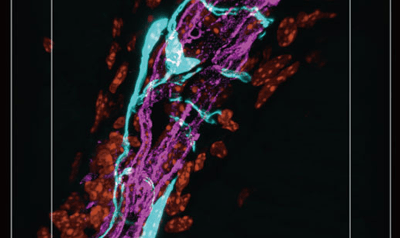
Understanding the vascular and neural projections to the knee joint is important for treating pain associated with knee arthropathy. However, researchers have been unable to fully observe the fine structures formed by sensory nerves and blood vessels throughout the knee joint due to the formation of complex neurovascular structures within a narrow area.
With the FV3000 microscope, these structures are clearly visible for the first time. Owing to its high transmission efficiency, the FLUOVIEW® FV3000 confocal laser scanning microscope enables bright, high-resolution imaging of fine structures while using low laser power, helping reduce photobleaching in the sample. Using this capability, researchers at the Keio University School of Medicine (Japan) were able to successfully image a complex 3D structure of sensory nerves and their surrounding vasculature in the tibial epiphysis. They observed that sensory nerves in the knee joint exist not only in the meniscus but also in the tibial epiphysis. These sensory nerves are entwined with surrounding blood vessels, and, together, the neurovascular structure penetrates a foramen in the tibial epiphysis. To learn more about how the FV3000 microscope facilitated this experiment, download the application note.