Meet the judges | BioTechniques Image Competition 2025
We are excited to announce our judging panel for the 2025 Image Competition.
After our submission window has closed, your images and supporting information will be sent to our judges. They will score each image based on scientific impact, image quality and wow factor, narrowing the pool of images down to the ten highest scorers. Then, we hand over to you; a public vote will follow, allowing you to choose which image is your favorite.
Michelle Itano – BioTechniques’ Editor-in-Chief
 Michelle Itano (left) is a cellular biophysicist, Assistant Professor of Cell Biology and Physiology and Director of the Neuroscience Microscopy Core at the University of North Carolina – Chapel Hill (NC, USA), where she develops and customizes state-of-the-art optical imaging and analysis applications for a wide range of scientific research. She utilizes innovative fluorescence microscopy methods — including dual selective plane illumination light-sheet imaging and iterative RNA in situ hybridization spatial distribution analysis in tissue — to investigate previously intractable questions in cell and neurobiology.
What is Michelle’s favorite scientific image from her research?
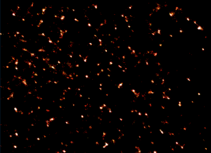 This is an image (right) of microdomains on the plasma membrane of human dendritic cells using super-resolution Blink microscopy, related to the research published in this paper. These nanodomains have a role in recognizing pathogens, like HIV-1. Michelle’s doctoral work focused on uncovering the nanoscale protein arrangement and mobility that enabled pathogen binding.
Joshua Rappoport – Executive Director, Research Infrastructure & Operations, Boston College (MA, USA)
 Joshua Z. Rappoport (left) received a bachelor’s degree in biology from Brown University (RI, USA) and then went on to earn a PhD from the Program in Mechanisms of Disease and Therapeutics at the Mount Sinai School of Medicine Graduate School of Biological Sciences of New York University (NY, USA). Following defense of his thesis, Joshua went on to perform postdoctoral work at The Rockefeller University in New York City (NY, USA) in the Laboratory of Cellular Biophysics. Subsequently he was recruited as a faculty member in the School of Biosciences at the University of Birmingham (UK).
In 2014, Joshua returned to the United States to become the Director of the Center for Advanced Microscopy and Nikon Imaging Center at the Northwestern University Feinberg School of Medicine (IL, USA), and a faculty member in the Department of Molecular and Cell Biology. Starting in March 2019, Joshua moved to Boston College (MA, USA), where he is the Executive Director, Research Infrastructure & Operations, working to assist with strategic organization and operations in the sciences.
What is Joshua’s favorite scientific image from his research?
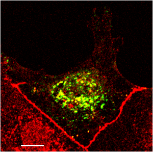
This image (right) is adapted from Figure 5 in the paper referenced below and was taken by first author Sarah (Fletcher) Dodds. It shows the edge of a monolayer of MDCK cells expressing rab5-GFP (green) that were wounded, allowed to migrate for 15 minutes and stained with anti-occludin (red) and then imaged by confocal laser scanning microscopy. It tells the story of the whole paper in one image.
José Manuel Martínez López – 2024 Image Competition winner
 José Manuel Martínez López (left) is an electromechanical engineer with a master’s in production engineering. His professional career started in drive systems design and finite element analysis, which has now evolved into a field that intersects with microscopy, science and photography. In 2006, José Manuel joined his sister to run their own company, Química Tech (Chihuahua, Mexico), where he specializes in microscopy.
When people ask José Manuel what he does for a living, he usually answers that he plays with microscopes and teaches others how to get the most out of their microscopes, particularly in the context of life sciences and photography.
What is José Manuel’s favorite scientific image from his work?
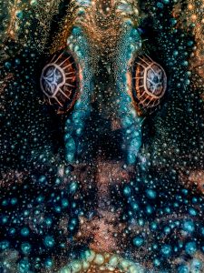 This photo (right) shows the dry eyes of a scorpion in autofluorescence after several hours under the microscope’s lens. It was captured using fluorescence microscopy, which is one of José Manuel’s favorite contrast techniques.
This image was part of our 2024 Image Competition Top 10.
Jasmine Hagan – Commissioning Editor of BioTechniques
 Jasmine (left) graduated from the University of Kent (UK) in 2021. Her interest in exploring new ways to remain connected with the scientific community, without direct involvement in laboratory work, led her to pursue a career in scientific publishing.
Jasmine joined Future Science Group in 2022 as a Commissioning Editor before moving into her current role at Taylor & Francis. In her role, she handles the day-to-day management of the BioTechniques journal. Her responsibilities include commissioning content for the journal and guiding authors through the publication process.
Her familiarity with BioTechniques’ scope and audience give her an important perspective on which images have potential to be our journal cover!