Go with your gut: exploring the microbiota–gut–brain connection
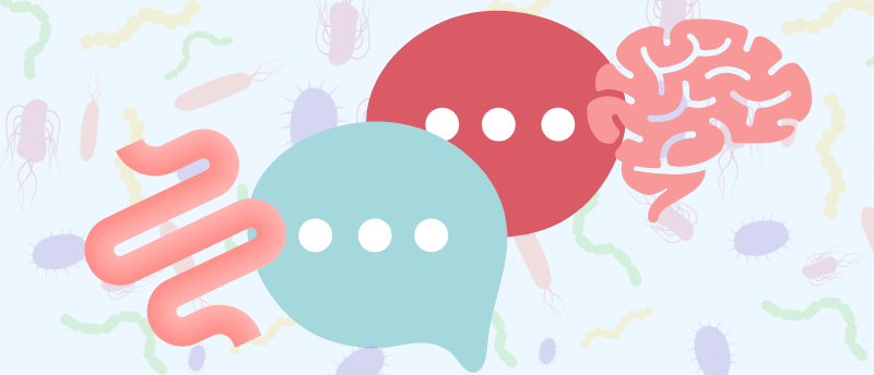
The far-flung organs of the gastrointestinal tract and the central nervous system are inextricably connected, and the growing body of scientific literature surrounding the ‘gut-brain axis’ is increasingly demonstrating the role of the microbiome in this two-way communication.
As our understanding of the role that microbiology plays in the maintenance of brain structure and function grows, the opportunity of exploiting these microbes therapeutically to combat neurological diseases is becoming more apparent. Here, we take a look at some recent preclinical advances in this area, from microbe-driven memory loss to the role of the gut microbiome in how exercise influences brain health and an antibiotic intervention for traumatic brain injury.
The gut microbiome can drive age-associated memory loss
Impaired gut–brain signaling and an aged microbiome are implicated in age-related cognitive decline in mice, research from the University of Pennsylvania (PA, USA) and the Arc Institute (CA, USA) has revealed. Providing some comfort, the team went on to demonstrate that various interventions show promise for returning the brain to a more youthful state.
Aging is accompanied by memory decline, which greatly affects the quality of life of a large number of older individuals. Brain-extrinsic factors, such as gastrointestinal signals, have been shown to influence age-associated memory loss, however, the underlying mechanisms remain largely unclear.
To investigate this knowledge gap, the team introduced gut microbial communities from aged mice into young recipients by co-housing 2-month and 18-month-old rodents. Frailty scoring revealed that their physical health was unaltered by microbiome equilibration, but the same couldn’t be said for their mental acuity.
In short-term novel object recognition and long-term spatial learning and memory tasks, young mice with older microbiomes performed poorly. Depleting their microbiomes using antibiotics restored memory performance in co-housed young mice, and even aged mice showed an improvement in cognitive ability.
The team performed metagenomic sequencing and proteomics on fecal content of a cohort of mice to characterize microbiome changes over the entire lifespan. This approach pinpointed Parabacteroides goldsteinii – a bacterium that produces medium-chain fatty acids – as a culprit behind this,
They also suggested a pathway for this association: bacteria like P. goldsteinii drive peripheral myeloid cell inflammation through production of medium-chain fatty acids, impairing the function of vagal sensory neurons and weakening the interoceptive signals received by the brain until, ultimately, hippocampal function declines.
Potential interventions, including the administration P. goldsteinii-targeting bacteriophages and stimulating the vagus nerve, reversed age-related memory deficits, the researchers later revealed. “These findings indicate a key role for interoceptive dysfunction in brain aging and suggest that interoceptomimetics that stimulate gut–brain communication may counteract age-associated cognitive decline,” they conclude.

PFAS versus the gut microbiome
Certain human gut microbes can absorb PFAS, offering potential protection against the harmful ‘forever chemicals’.
Antibiotics remodel gut microbiome to aid recovery from traumatic brain injury
Antibiotic treatment appears to significantly reduce neuroinflammation following traumatic brain injury by altering the gut microbiome in animal models, a new study suggests.
The microbiota–gut–brain axis plays a role in regulating brain function, and disruption to gut bacterial composition may impair its protective function and result in neuroinflammation. Conversely, traumatic brain injury can cause microbiome dysbiosis; however, the effects of short-term antibiotic treatment on these processes are poorly understood.
To address this, a research collaboration between Houston Methodist Research Institute and Rice University (both TX, USA), induced brain injuries in male mice before administering a brief course of oral antibiotics. Some mice received a single controlled cortical impact, while for others this was repeated. The effects of antibiotic treatment were assessed via fecal microbiome amplicon and metagenomic sequencing, metabolite profiling and neuropathological analyses.
The findings revealed that antibiotic administration altered microbial community composition, with more pronounced shifts following repeated brain injury. However, despite this disruption, the treatment also seemed to decrease lesion size, diminish neuroinflammatory responses and limit cell death, suggesting that microbial depletion may contribute to neuroprotection.
Long-read metagenomic sequencing identified two beneficial bacteria, Parasutterella excrementihominis and Lactobacillus johnsonii, which persisted after antibiotic treatment and may be key to repairing damage.
Taken together, these results suggest that antibiotics can reduce brain damage after injury and “support a gut–brain mechanism in which microbiome changes influence peripheral immunity and, in turn, neuroinflammation after [traumatic brain injury],” study lead Sonia Villapol (Houston Methodist Research Institute) concluded.
Next, the team hopes to bioengineer P. excrementihominis and L. johnsonii to develop precision therapies to reduce neuroinflammation.
Light therapy shows promise for treating traumatic brain injury
Near-infrared light therapy has reduced inflammation and improved recovery in a mild traumatic brain injury rat model.
High-fat diet drives gut microbiome bacteria into the brain
Researchers from Emory University (GA, USA) have discovered that bacteria can translocate directly from the gut to the brain when mice are fed a high-fat diet that causes alterations in gut microbiome composition, with potential implications for neurological health.
A growing body of research has hinted at a potential link between gut dysbiosis and neurodegenerative or neurodevelopmental conditions such as Alzheimer’s disease, Parkinson’s disease and autism spectrum disorder. However, currently known mechanisms fail to explain how the gut microbiome might directly affect the brain to cause pathologies associated with neurological diseases.
In search of an explanation, the team fed a group of germ-free mice a high-fat ‘Paigen diet’, with 45% carbohydrate and 35% fat content, for 9 days. In humans, this diet is associated with an altered gut microbiome and intestinal permeability or a ‘leaky gut’.
In mice following this diet, bacteria were detected in the vagus nerve, but not in any other systemic sites or blood, and right cervical vagotomy reduced bacterial burden in the brain, further implicating the vagus nerve as a channel for bacterial translocation from the gut to the brain.
When the scientists administered antibiotics to mice for 3 days, they noticed that it impacted the composition of the gut microbiome and changed the bacteria that localized to the brain in mice fed a Paigen diet.
They then fed the mice Enterobacter cloacae engineered with a DNA sequence not normally found in these bacteria in nature. When the mice were also fed the Paigen diet, E. cloacae was detected in the gut and brain.
The team also identified bacteria in the brain in murine models of Alzheimer’s, Parkinson’s and autism spectrum disorder fed a standard diet.
“This research highlights the need for further study into how dietary shifts have a huge influence on human behavior and neurological health,” commented Arash Grakoui, co-principal investigator of the study.

The Mediterranean diet and cognition: is the gut microbiome the key?
Investigations of the Mediterranean diet, the gut microbiome and cognition have revealed intriguing associations between the three…
Exercise reshapes the gut microbiome to influence the brain
We have long known that exercise can have beneficial effects on mood and memory. One pathway through which it is believed to exert these effects is via the gut microbiome, which produces metabolites that can influence brain function. However, we do not yet know which exercise-induced alterations in gut microbiota are associated with alterations in systemic metabolites that may affect the brain.
Keen to rectify this, a team of researchers from University College Cork (Ireland), gave rats free access to a running wheel for 8 weeks and investigated exercise-driven changes in their gut microbiome composition and serum metabolite profile, as well as the potential contribution of these changes to hippocampal processes implicated in memory and mood.
Using 16S rRNA gene amplicon sequencing analysis of rat fecal material, the researchers revealed that exercise modified the gut microbiota diversity. Specifically, it increased microbial dominance: the relative abundance of the most prevalent species in a microbiome. Exercise also decreased the relative abundance of two bacterial genera, Clostridium and Alistipes, both of which are associated with tryptophan metabolism.
The researchers next performed untargeted serum metabolomics, which revealed that exercise enhanced tryptophan metabolism, with a notable enhancement of the serotonin catabolite 5-hydroxytryptophol identified.
Pathway analysis of the gut–brain modules revealed that tryptophan metabolism was enhanced by exercise. Moreover, exercise decreased hippocampal expression of the aryl hydrocarbon receptor, a mediator of the effects of tryptophan-metabolizing gut microbes on neuronal function. This points to a pathway whereby exercise modulates gut microbes associated with systemic tryptophan metabolism, which may then exert beneficial effects on the brain.
“What struck us most was the convergence of evidence across multiple levels of analysis,” remarked Yvonne M. Nolan, corresponding author of the study. “Each finding alone would be noteworthy. Together, they suggest a coherent biological pathway through which the gut microbiota may mediate the beneficial effects of exercise on brain regions critical for memory.”