Harnessing mass spectrometry imaging to generate lipid maps
Original story from Okayama University (Japan).
A new microfluidic method preserves worm anatomy to link lipid signals with body structure using mass spectrometry imaging.
Understanding how fat molecules are distributed and function in living organisms is key to uncovering mechanisms of aging, disease and metabolism. Caenorhabditis elegans, a transparent nematode, is a widely used model for studying fat storage due to its genetic similarity to humans and well-defined anatomy. However, visualizing lipids at high resolution in such a small organism has posed a major technical challenge.
A research team at Okayama University (Japan) led by Masazumi Fujiwara and Sara Mandic, in collaboration with Ron M. A. Heeren (Maastricht University, Netherlands), has developed a new microfluidics-based workflow that enables high-resolution, 3D lipid imaging in C. elegans.
The team combined matrix-assisted laser desorption/ionization mass-spectrometry imaging (MALDI-MSI) with conventional lipid staining techniques to map both the identity and location of lipid molecules inside the worm. To preserve internal structures, young adult nematodes were aligned and immobilized on a custom-designed microfluidic chip, embedded in a gelatin–carboxymethyl cellulose mixture, sectioned using a cryotome and analyzed through MALDI-MSI. Each section was also subjected to Oil Red O staining, which highlights neutral fats, to confirm and complement the imaging results. “This is the first time we’ve been able to map lipid distributions in C. elegans with such spatial resolution while preserving internal structures,” explained Mandic.
Conventional lipid analysis techniques often involve trade-offs – either staining lipids without identifying them or measuring them without preserving spatial information. This new method does both: it detects specific lipid molecules and shows where they are located inside the body. The ability to retain the internal anatomy of the worm during sample preparation is key to understanding how lipids behave in different tissues. “Our technique gives researchers a reliable way to study fat dynamics in specific tissues of a single nematode,” commented Mandic.
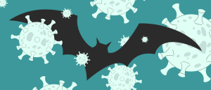 Bat organoid platform reveals more about zoonotic viruses
Engineered mini organs from the most common bat species have been developed to study infections by key viruses, including COVID-19 and influenza.
Using their method, the team identified several lipids that clustered in distinct anatomical regions, including the pharynx, intestine and reproductive system. For instance, one lipid linked to cholesterol metabolism was found primarily in the pharynx and anterior intestine, suggesting a potential role in nutrient absorption. These findings, made possible by the method’s structural preservation, provide valuable insight into how fat molecules are organized and function across different parts of the worm.
The researchers also extended their analysis beyond two-dimensional imaging. By aligning and stacking consecutive tissue slices, they created three-dimensional reconstructions of individual nematodes, offering a full-body view of lipid distribution with remarkable anatomical detail. This approach allowed them to visualize how lipids are arranged throughout the entire ~1 mm-long organism – something that previously had not been achieved at this level of precision. Importantly, the method was shown to be highly reproducible. Variations between individual nematodes were greater than any technical inconsistencies, demonstrating the accuracy and robustness of the workflow. “This method allows us to see not just what lipids are present, but exactly where they are inside the body – whether in the intestine, pharynx or embryos,” added Mandic.
Because C. elegans share many fundamental biological pathways with humans, this technique has wide-reaching implications for biomedical research. It enables the study of lipid behavior in response to genetic mutations, environmental stress, drug treatments and aging – all key factors in human health and disease. The team now plans to apply this workflow to various C. elegans strains, including those carrying disease-related mutations, and to integrate the technique with lipid quantification tools.
“Our work opens the door to visualizing lipid biology in an entirely new way – one that’s precise, reproducible and rich in detail,” concluded Mandic. Altogether, this study equips researchers with a powerful tool for examining fat metabolism at the organ-specific level in C. elegans, paving the way for deeper insights into aging, metabolic disorders and disease mechanisms.
This article has been republished from the following materials. Material may have been edited for length and house style. For further information, please contact the cited source. Our press release publishing policy can be accessed here.