Could chronic pain be a thing of the past? Uncovering the molecular signature of ‘sleeping’ nociceptors
Original story from The University of Texas at Dallas (TX, USA).
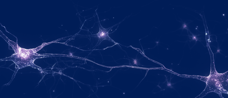
The molecular signature of key sensory neurons that signal neuropathic pain has been uncovered, revealing a potential pathway for relieving chronic pain.
Researchers from The University of Texas at Dallas (TX, USA) and their international colleagues have determined the molecular signature of human sleeping — or silent — nociceptors: sensory neurons that are unresponsive to touch or pressure yet are key culprits in neuropathic pain. The findings suggest a potential pathway for finding drug targets to relieve chronic pain, shared Ted Price, Ashbel Smith Professor of neuroscience in the School of Behavioral and Brain Sciences and a co-author of the study.
“We know from direct human physiological evidence that these cells are important in neuropathic pain,” explained Price, director of the UT Dallas Center for Advanced Pain Studies. “Now we can identify them at the gene-expression level with an astonishing degree of detail. This will allow researchers to start working on targets to manipulate those cells, which could bring about very exciting developments in the future.”
Sleeping nociceptors are a distinct class of sensory neurons that can become spontaneously active to cause persistent pain without an evident stimulus. This makes them essential components of the neuropathic pain suffered by approximately 20% of American adults.
The cell bodies of sleeping nociceptors are located in the dorsal root ganglia, nerve cells clustered near the base of the spine that relay sensory signals from the peripheral nervous system to the central nervous system. The axons and dendrites that protrude from each cell and carry electrical signals are long and connect to the skin. Though the functional properties of these fibers have long been known, their distinctive molecular characteristics remained unclear.
“Active sleeping nociceptors have been found in people with diabetic neuropathy, postherpetic neuralgia and fibromyalgia,” Price commented. “There are also scores of neuropathic pain studies that found no cause. If you were going to highlight any type of sensory neuron as the biggest culprit for the spontaneous, shooting pain that neuropathy patients have, it’s these sleeping nociceptors.”
 ‘Silent’ X chromosome reduces disease severity in the female brain
X chromosome reactivation contributes to reducing the signs of disease in the female brain.
Led by corresponding author Angelika Lampert, professor of neurophysiology at RWTH Aachen University (Germany), the researchers used high-resolution recordings of electrical activity of individual neurons alongside techniques that read the genetic activity of the neurons to identify sleeping nociceptors among the broader nerve population.
To discover the distinctive molecular profile of the neurons, the German researchers first used isolated dorsal root ganglia from pigs because sleeping nociceptors in porcine skin closely resemble those found in humans. Cross-species analyses confirmed that the same molecular markers are present in pig and human sensory neurons. They are both characterized in part by the presence of the oncostatin M receptor on the cell and the neuropeptide somatostatin, which suppresses the release of various hormones. “Our work establishes a new conceptual framework for understanding the emergence of neuropathic pain at the molecular level, opening concrete perspectives for the development of targeted therapies,” Lampert shared.
Co-corresponding author Shreejoy Tripathy, associate professor of psychiatry at the University of Toronto (Canada), led the bioinformatic integration of the patch-sequencing data, linking the neurons’ functional properties with their gene expression profiles to identify neuronal subtypes whose activity resembled that of sleeping nociceptors. “This collaboration produced a Rosetta stone for pain research, matching the electrical fingerprint of sleeping nociceptors to a specific genetic signature,” Tripathy commented.
Co-second author Marisol Mancilla Moreno, a cognition and neuroscience doctoral student in Price’s lab, led the portion of the project revolving around spatial sequencing, which is used to show which genes are particularly active in different cell types.
“We now hope to start a drug discovery project to try to silence these cells,” Price explained. “The dataset we’ve generated on the molecular characteristics of these cells is so comprehensive that the modeling is going to be extremely instructive.”
Lampert concluded: “When you realize the kind of team you need to assemble to answer these questions, you turn to the best people you can find at each portion of the project — collaboration partners with whom you know you can push projects and with whom you enjoy working. The success of the study relies on the close integration of specialized centers, like UT Dallas. This work demonstrates the power of interdisciplinary and international cooperation.”
This article has been republished from the following materials. Material may have been edited for length and house style. For further information, please contact the cited source. Our press release publishing policy can be accessed here.