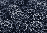
Dual-mode bioimaging with fluorescent nanoparticles

Published in ACS Nano, researchers from the KTH Royal Institute of Technology (Stockholm, Sweden) have described engineered nanoparticles that combine X-ray and optical fluorescence properties. The nanoparticles – which are coated in silica and have a fluorophore conjugated to their silica coatings – could represent effective macro and microscopic imaging tools.
Read the full story on our sister site, Bioanalysis Zone, now >>>
“This unique design of nanoparticles paves the way for in vivo tumor diagnostics, using X-ray fluorescence computed tomography,”
– Muhammet Toprak, contributing study author and Professor of Materials Chemistry at the KTH Royal Institute of Technology.
You may also be interested in…
- Using nanoparticle ‘pseudo-virions’ to screen for potential COVID-19 therapeutics: an interview with Kirill Gorshkov
- Mirror SELFI tracks the position of nanoparticles by generating self-interference patterns
- Peek behind the paper: superparamagnetic iron-oxide nanoparticles as potential theranostic agents