Imaging technology allows 3D visualization of nanoscale structures
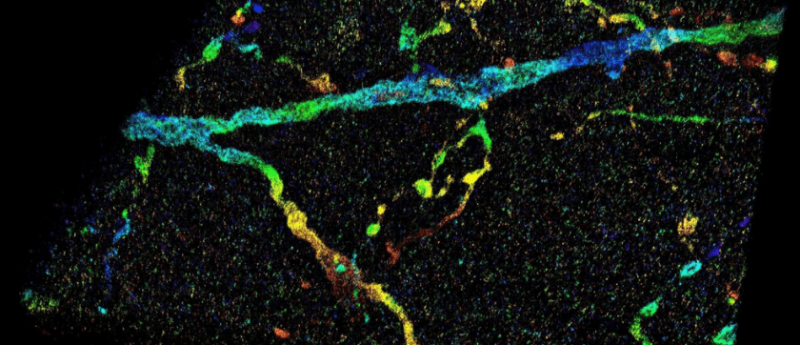
Researchers from Purdue University (IN, USA) have developed a new imaging technology to overcome previous issues associated with the resolving capabilities of super-resolution fluorescence microscopy for observing inside whole cell or tissue samples. Such technology will vitally advance the study and understanding of structural analyses for cancers and other diseases. Previously, the challenges associated with super-resolution fluorescence microscope imaging revolved around the light signals emitted from molecules inside a specimen. These travel through different parts of the cell or tissue structure at different speeds and results in deviations, which deteriorate the image. The team’s novel technology overcomes this issue by...
To view this content, please register now for access
Join our member community for FREE to access a collection of journal and online-only features, including:
- Exclusive access to educational videos, eBooks and insights into top BioTechniques journal articles
- The latest news and journal updates delivered straight to your inbox when you want it
- Personalized recommendations for the latest member-exclusive podcasts, interviews and expert opinions
- Priority registration to webinars, panel discussions and events
- Access to competitions and journal publication discounts, including 10% off open access fees when you sign up today!