The holy grail of neuroimaging: the revolutionary new sensor
The development of a novel sensor allows electrical brain activity to be measured for a longer period of time and deeper within the brain than previously possible.
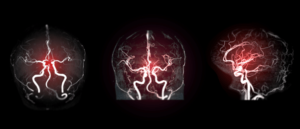
The electrical activity displayed by the brain is critical in enabling both our cognitive and our physical abilities. New insights into understanding how this electrical brain activity operates could unlock potential treatment avenues for a variety of neurological related disorders. Current noninvasive technologies are limited to observing activity at the surface of the brain, missing key brain signals. Now, a collaborative study led by a team of researchers from Baylor College of Medicine (TX, US) reports a novel sensor able to image brain activity deeper in the brain and for up to 40 minutes.
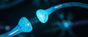
Measuring electrical activity within the brain is not a new phenomenon. In fact, there are many existing technologies that have been used for this purpose; however, each comes with its own pitfalls. François St-Pierre (Baylor College of Medicine and Rice University; TX, USA) commented: “For example, electrodes can record very fast activity, but they cannot tell what type of cells they are listening to.” The use of fluorescent proteins, which respond to fluctuations in calcium levels associated with electrical activity, is also common in this field and can be monitored with a two-photon microscope. “This kind of sensor is excellent to determine which neurons are active and which are not. However, they are very slow. They measure voltage changes indirectly, thereby missing a lot of key signals,” stated St-Pierre.
The research team, therefore, aimed to combine the best elements of the existing technologies, to develop a sensor capable of measuring electrical brain signals whilst monitoring any associated changes in specific cells. St-Pierre commented on the novelty of this research: “It has been challenging to figure out how to noninvasively observe the millisecond-fast electrical activity in individual neurons of specific cell types in animals carrying on an activity. To be able to do this has been the holy grail of neuroimaging.”
 Expansion revealing microscopy exposes unseen cellular nanostructures
Expansion revealing microscopy expands tissue and cell samples to improve the resolution of fluorescence imaging.
The technique is underpinned by engineered fluorescent proteins, termed voltage indicators. An automated system was used to optimize these fluorescent voltage indicators for two-photon microscopy, which is commonly used for noninvasive deep-tissue neuroimaging. Zhuoche (Harry) Lui (Rice University) reported: “Using this system, we tested thousands of indicator variants and identified JEDI-2P, which is faster, brighter and more sensitive and photostable than its predecessors.”
This new technique overcomes previous challenges, as reported by Xiaoyu (Helen) Lu (Rice University): “First, it allows us to follow electrical activity in a living animal for as long as 40 minutes instead of at most a few minutes. Second, we can image spikes of electrical activity with a temporal resolution of about one millisecond, and third we can image individual cells deeper in the brain because our indicator is bright and produces large signals in response to brain activity.” In fact, this sensor allows neuroscientists to non-invasively measure electrical signals within the cortex, no longer limiting observations to the surface of the brain.
The abilities of this technology permit the noninvasive monitoring of brain signals in living animals, paving the way for understanding further how the brain functions, in both a healthy and diseased state.