The neuroscience behind perceiving illusions
Original story from the Allen Institute (WA, USA).
Researchers investigate how mice process illusions, highlighting the neural circuitry involved in seeing and perception.
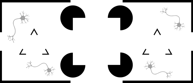
An illusion is when we see and perceive an object that doesn’t match the sensory input that reaches our eyes. In the case of the Kanizsa square, the sensory input is four Pac Man-like black figures; however, what we see or perceive is a white square – i.e., the illusion.
In a new study, researchers from the University of California, Berkeley (CA, USA), worked with teams at the Allen Institute (WA, USA) to identify the key neural circuit and cell type that plays a pivotal role in detecting these illusions – more specifically, their outer edges or ‘contours’ – and how this circuit works.
Hyeyoung Shin (now with Seoul University, South Korea), Hillel Adesnik (UC Berkeley) and their team discovered a special group of cells called IC–encoder neurons that tell the brain to see things that aren’t really there as part of a process called recurrent pattern completion.
“Because IC–encoder neurons have this unique capacity to drive pattern completion, we think that they might have specialized synaptic output connectivity that allows them to recreate this pattern in a very effective manner,” explained Shin. “We also know that they receive top-down inputs from higher visual areas. The representation of the illusion arises in higher visual areas first and then gets fed back to the primary visual cortex; and when that information is fed back, it’s received by these IC–encoders in the primary visual cortex.” This is like a manager telling an entry level worker to complete a task; instructions come from a higher-level and are then executed by lower-level staff. In this case, the instruction would be to see or perceive something that isn’t really there.
 The bonds that grow stronger the more they’re pulled apart
AI analysis uncovers why certain protein interactions behave like a finger trap, gripping tighter the harder they’re pulled.
In the context of the brain and vision – referring to the Kanizsa square mentioned earlier – higher levels of the brain interpret the image as a square and then tell the lower-level visual cortex to ‘see a square’ even though the visual stimulus consists of four semi-complete black circles.
Shin, Adesnik and their team made the discovery by observing the electrical brain activity patterns of mice when they were shown illusory images like the Kanizsa triangle. They then shot beams of light at the IC-encoder neurons, in a process called two-photon holographic optogenetics, when there was no illusory image present. When this happened, they noticed that even in the absence of an illusion, IC-encoder neurons triggered the same brain activity patterns that exist when an illusory image was present. They successfully emulated the same brain activity by stimulating these specialized neurons.
The findings shed light on how the visual system and perception work in the brain and have implications for diseases where this system malfunctions. “In certain diseases you have patterns of activity that emerge in your brain that are abnormal, and in schizophrenia these are related to object representations that pop up randomly,” commented Jerome Lecoq, associate investigator at the Allen Institute. “If you don’t understand how those objects are formed and a collective set of cells work together to make those representations emerge, you’re not going to be able to treat it; so understanding which cells and in which layer this activity occurs is helpful.”
Researchers with the Allen Institute’s OpenScope program – which allows external scientists to propose experiments that can be done using the institute’s cutting-edge tools and equipment – conducted some of the experiments that were part of this study. Their work showed for the first time that the brain activity ‘feedback loop’ from higher-order levels of the brain communicating to lower visual areas (where the IC-encoders neurons were) happens in mice.
This article has been republished from the following materials. Material may have been edited for length and house style. For further information, please contact the cited source. Our press release publishing policy can be accessed here.