3D image analysis — live/dead cell assay of spheroids
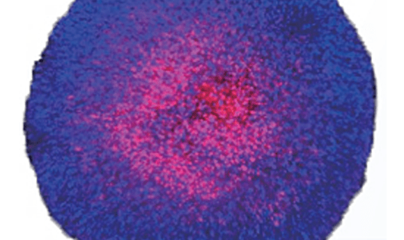
The distance from the tumor surface to the core has a diffusion gradient of oxygen or nutrients, and this severe environment causes resting cells or dead cells deep inside the tumor tissue. A spheroid model is expected to recapitulate tumor microenvironments. In this study, confocal images of fluorescent stained cancer spheroids, treated with a drug, fixed, and made transparent, were subjected to 3D quantitative analysis using Olympus NoviSight™ software.
The study demonstrates that drug-independent and drug-dependent cell death can be confirmed using a clearing reagent and NoviSight software. To learn more about NoviSight 3D high content analysis for spheroid models, download the application note.
You might also be interested in...