Cryo-electron tomography contributes to our understanding of bacterial interactions with their environment
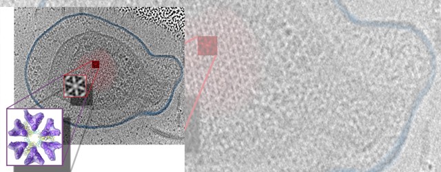
How are microbes able to actively seek out their preferred environmental niches, how can they effectively evade toxins, and how can they adapt to thrive in their changing environments? In order to gain insight into the structure and function of the macromolecular complexes involved in these behaviors, we use cryo-electron tomography (Cryo-ET). This technique allows us to directly study microbes in their native state at resolutions capable of visualizing individual proteins.
What will you learn?
- How Cryo-ET can help understand the macromolecular machines bacteria use to sense and interact with their environment
- How Cryo-ET (a cryo-electron microscopy method) works
Who may this interest?
- Academic scientists (universities, research institutes and hospitals)
- Imaging core facilities
Speaker

Ariane Briegel
Professor of Ultrastructural Biology, Co-Director NeCEN
Leiden University
Ariane Briegel is a professor at Leiden University (The Netherlands). She has over 15 years of experience using cryo-electron microscopy to study bacterial and archaeal ultrastructure. The Briegel laboratory focuses on investigating how microbes sense and respond to their environment. In order to gain insight into the structure and function of the molecular complexes involved in these behaviors, the lab uses electron cryotomography and correlative microscopy methods.
This webinar was recorded on 13th December 2019
In association with
![]()