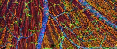
Multiphoton microscopy for deep tissue penetration
Since scientists first glimpsed microscopic organisms, they have been searching for ways to probe ever smaller details in greater resolution. Monumental scientific and engineering achievements pushed beyond resolution limits have enabled researchers to image a wide range of biological creatures, tissues, cells, proteins, and processes. Now, many researchers want to explore cells and their functions in their native environments.
In this webinar, Chris Xu will present his latest work using multiphoton microscopy to peer deep inside the brain. He’ll review the need for such technology, the challenges he and his team overcame in developing their approach, and demonstrate what can be accomplished with deep imaging.
In this webinar, you will learn:
- Why researchers need deep tissue imaging
- The challenges involved with peering deeper inside tissues
- How tissue preparation methods can help resolve those challenges
- Microscopy techniques and instrumentation for achieving deep imaging
- Examples of recent explorations deep within the mouse brain
In addition, Dr. Xu will discuss the applications best suited for deep imaging using multiphoton microscopy, the equipment and steps needed to implement the approach in your own lab, and how to establish a collaboration to pursue your own deep imaging research questions.
Featured speaker:
Chris Xu
Cornell University
Chris Xu is a professor in the School of Applied and Engineering Physics at Cornell University, the Mong Family Foundation Director of Cornell Neurotech–Engineering, and the director of Cornell NeuroNex Hub, an NSF funded center for developing neurotechnology. His research focuses on fiber optics and biomedical imaging, including multiphoton microscopy.
Sponsored by:
![]()