Panel Discussion: Imaging in neuroscience
Imaging methods help neuroscientists understand the structure and function of the central nervous system, from cell-to-cell interactions and signal transduction to regional brain connectivity and differences between varying functional areas. Understanding these gives us insight into various functions, such as speech and memory, and how these are impacted in neurological disorders, including dementia and Alzheimer’s, which have become more common as the population ages.
In this Panel Discussion for our Spotlight on imaging in neuroscience, we’ll explore various developments and hot topics in the field, including technologies such as spatial transcriptomics, with experts in the field.
What will you learn?
- The latest developments in neuroimaging
- The use of single-cell and spatial analyses in neuroscience
- Customizing imaging equipment
Who may this interest?
- Anyone working in neuroimaging
- Neuroscientists
- People wanting to learn more about neuroimaging
Panellists

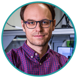
Robert Prevedel
Group Leader
European Molecular Biology Laboratory
Robert Prevedel is a researcher at the European Molecular Biology Laboratory (EMBL; Heidelberg, Germany), whose lab focuses on developing light microscopy-based techniques for a variety of applications, including neuroimaging techniques that capture brain activity and structure in different model organisms, focusing especially on mouse models.

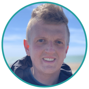
Harry Fu
Assistant Professor
The Ohio State University
Harry Fu is an Assistant Professor in the Department of Neuroscience at The Ohio State University College of Medicine (OH, USA).
Harry’s research focuses on understanding which subtypes of neurons are vulnerable to tau pathology in early Alzheimer’s disease and other tauopathies as well as the molecular and cellular mechanisms underlying the selective neuronal vulnerability.
He utilizes a multidisciplinary approach that combines neuropathology, mouse genetics, neurobehavioral tests, confocal and light-sheet microscopy, molecular and cell biological approaches together with single cell RNA-seq and spatial transcriptomic analysis.

Filip Janiak
Research Fellow
University of Sussex
Filip Janiak has a MSc and PhD in the field of solid-state physics, obtained from Wroclaw University of Science and Technology (Poland).
After his PhD, Filip was at NeuroElectronics Research Flanders (Leuven, Belgium) for a short time, before moving to Kavli Institute for System Neuroscience (Trondheim, Norway), where he was a postdoc in Emre Yaksi’s lab.
Since 2017, Filip has been a Research Fellow in Tom Baden’s lab at the University of Sussex (UK). His research focuses on in-vivo 2-photon imaging of neural activity in the eye and brain.

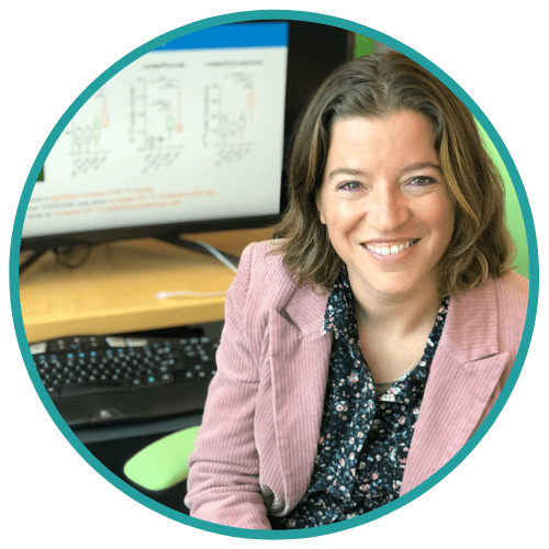
Myriam Chaumeil
Associate Professor
University of California, San Francisco
Myriam Chaumeil is an Associate Professor in Residence in the departments of Physical Therapy & Rehabilitation Science, and Radiology & Biomedical Imaging at the University of California, San Francisco (CA, USA). She is also a research group leader in Metabolic Imaging of Neurological Disorders.
Myriam’s research focuses on developing and validating magnetic resonance-based imaging and spectroscopy methods for in vivo measurement of brain metabolism, in physiological and pathological conditions, in preclinical models and in patients. She has experience in studies of Huntington’s disease, multiple sclerosis, Alzheimer’s disease, traumatic brain injury, cerebral small vascular diseases and CNS lymphoma.
This webinar was recorded on Thursday 20th October 2022
