Researchers identify a promising therapeutic target for sepsis
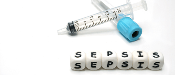
Autophagy can alleviate sepsis by restoring intestinal barrier function, and now researchers have identified a potential therapeutic target to promote this process.
Sepsis is one of the most serious disease complications that can occur in the intensive care unit, resulting in life-threatening organ dysfunction, and is exacerbated by the disruption of the intestinal barrier. Researchers at The Yijishan Hospital of Wannan Medical College (Anhui, China) have demonstrated how inducing autophagy, the process of cellular break down and protein destruction, using an immunosuppressant reduced disruption to the intestinal barrier.
The researchers used an in vivo model of sepsis by subjecting mice to cecal ligation and puncture (CLP), a procedure that induces sepsis by releasing fecal material into the peritoneal cavity. The mice were then treated with a common immunosuppressant, rapamycin. This treatment reduced intestinal epithelial cell death and restored the disrupted intestinal barrier, showing that autophagy provides some level of protection against sepsis-induced intestinal barrier dysfunction.
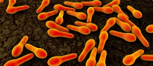 Stop: hammer time! Identifying the lethal mechanism of Clostridium septicum
Bacteria that kill cells through a hammer-like mechanism may soon be stopped in their tracks.
“Despite the increased understanding of sepsis pathophysiology and the application of advanced clinical treatments, sepsis remains a major cause of health loss worldwide with a high health-related burden,” explained Wei-Hua Lu, the lead author of the study. Lu added that the function of the mammalian target of rapamycin (mTOR) and polo-like kinase 1 (PLK1) in sepsis was not well understood and investigated this further.
The researchers wanted to investigate whether the protective ability of autophagy relies on PLK1 by modifying mice with the PLK1 gene to overexpress this protein and subjecting them to CLP. Autophagy occurred, and cell death through apoptosis was alleviated. Some mice were treated with chloroquine to inhibit autophagy, and the protective benefits of PLK1 were no longer observed. This indicates that PLK1 promotes intestinal autophagy and provides protection against sepsis-induced barrier dysfunction in doing so.
The interplay between PLK1 and mTOR was then studied in vitro using a model of human colonic epithelial cells. The results showed that PLK1 physically interacts with mTOR and participated in a reciprocal regulatory crosstalk in intestinal cells during sepsis.
“The reciprocal regulation of the PLK–mTOR axis is crucial in sepsis-induced intestinal barrier dysfunction,” explained Lu. “These findings indicate that the PLK1–mTOR axis may be a promising therapeutic target for treatment of sepsis.”

