New way to diagnose ovarian cancer with extracellular vesicle membrane proteins

Membrane proteins expressed on ovarian cancer-derived extracellular vesicles (EVs) are shown to be suitable biomarkers for ovarian cancer detection.
Researchers at Nagoya University (Japan) identified three previously unknown membrane proteins on EVs derived from ovarian cancer that could be suitable biomarkers for its early detection. They also developed a technology to capture the proteins from blood samples and demonstrated its clinical utility as a detection method.
Ovarian cancer is challenging to detect in its early stages when it is most easily treated. This is because ovaries are located deep within the abdominal cavity making it difficult to feel a lump or spot abnormalities in the area. Additionally, there is no single test to diagnose ovarian cancer, and therefore, finding new biomarkers is vital to improve survival rates.
Tumors release EVs, which have recently been explored for cancer detection, especially small proteins called exosomes. As these proteins are outside of cancer cells, they can be isolated from body fluids like blood, urine and saliva. However, EVs in body fluid are fairly heterogenous, hindering the identification of disease-specific EV markers.
 A wound-healing bioink accelerates skin repair
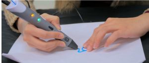
Researchers have developed a bioactive hydrogel ink containing macrophage-derived extracellular vesicles (EVs) that enhances cutaneous wound healing and can be administered in situ using a 3D-printing pen.
The researchers of this study focused their efforts on high-grade serous ovarian carcinoma (HGSOC), the most common type of ovarian cancer, and extracted small and medium/large EVs from cell lines and patient serum. They then performed shotgun proteomics using liquid chromatography–tandem mass spectrometry (LC–MS/MS) to decipher their protein profiles.
“The validation steps for the identified proteins were tough because we had to try a lot of antibodies before we found a good target,” commented lead author Akira Yokoi. “As a result, it became clear that the small and medium/large EVs are loaded with clearly different molecules. Further investigation revealed that small EVs are more suitable biomarkers than the medium and large type.”
Continued analysis revealed three membrane proteins in small EVs that are associated with HGSOC – FRα, Claudin-3 and TACSTD2.
Having identified possible protein biomarkers, the researchers collaborated with nanowire specialist Takao Yasui to develop a method to capture the EVs. They developed a novel isolation method with polyketone chain-coated nanowires (pNWs) to separate exosomes from blood samples.
“The results of this research suggest that these diagnostic biomarkers can be used as predictive markers for specific therapies,” concluded Yokoi. “Our results allow doctors to optimize their therapeutic strategy for ovarian cancer, therefore, they may be useful for realizing personalized medicine.”