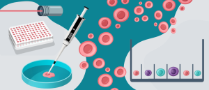
Advancing drug discovery with cell line development: past, present and future

Cell line development is an essential step in the manufacture of biologics:drugs derived from living organisms, such as monoclonal antibodies, recombinant proteins and vaccines. In addition to this, cell lines are vital for drug screening, toxicity testing, disease modeling and investigating gene function; in a nutshell, they represent a crucial tool in the drug discovery and development pipeline.
Here, we explore the process of cell line development and its contributions to drug discovery, as well as how the field has evolved over the years and the technologies that are helping it to advance.
The history of cell line development
The foundations of cell line development were laid in the late 19th and early 20th century, when the practice of culturing cells first took off. In the 1880s, Wilhelm Roux, of the University of Halle (Germany), demonstrated that it was possible to keep cells alive in a saline solution. Then, in 1906, Ross Granville Harrison (Johns Hopkins University; MD, USA) cultivated frog nerve cells in test tubes containing a liquid medium composed of blood clots, saline and agar. He was also instrumental in developing the ‘hanging drop’ technique, which is still used in modern cell culture research, and involves culturing cells within a droplet of plasma on the underside of a glass slide.
The decades that followed heralded a number of breakthroughs, spurred on by the introduction of aseptic technique. In 1911, for example, Alexis Carrel of the Rockefeller Institute (NY, USA) described the first 3D cell culture method when he cultivated tissue fragments on silk threads saturated with plasma. Carrel was also responsible for the isolation and cultivation of one of the first immortalized cell lines derived from chicken embryonic hearts.
In 1943, a mouse fibroblast cell line called ‘L cells’ was established by William Earle at the National Cancer Institute (MD, USA). This was followed by the first human cell line in 1951, created by George Gey (Johns Hopkins University). These ‘HeLa’ cells are derived from Henrietta Lacks’ cervical cancer cells, without her consent, and have been used in countless scientific discoveries ever since, including developing the first polio vaccine in 1953.
Six years later, in 1957, the first Chinese hamster ovary (CHO) cell line was established when researchers at the University of Colorado Medical School (CO, USA) extracted epithelial cells from the ovary of a female Chinese hamster. CHOs were involved in the first mammalian-expressed recombinant therapeutic to gain FDA approval (1987) – human tissue plasminogen activator – and are now a staple of biotherapeutic production.
Another prominent cell line, HEK-293, was generated in 1973 at Leiden University (Netherlands) when Alex Van der Eb and Frank Graham transfected normal healthy human embryonic kidney cells with adenovirus 5 DNA. This line and its many subtypes are often used to produce therapeutic proteins and viruses for gene therapy, and were involved in the manufacture of adenoviral vectored COVID-19 vaccines.

Cell line development: pitfalls, challenges and solutions
How technological advances are helping to overcome challenges achieving monoclonality and assessing productivity to streamline and de-risk cell line development.
Stages of cell line development
To establish a stable cell line for advancing drug discovery and development, researchers must first determine the most suitable cell line. Creating new cell lines can be challenging, so scientists often modify existing ones, like CHO cells, HeLa cells or human embryonic kidney-derived epithelial (HEK293) cells, instead of starting from scratch.
The first step of cell line development is gene cloning and transfection. Cells of the selected line are transfected with an engineered plasmid encoding the desired protein. The plasmid enters the nucleus and integrates into the host DNA.
Then, cells are isolated and expanded to create clonal populations. Stable clones with high expression levels of the protein of interest are selected, and the monoclonality of the cell population is verified. Once this screening step is complete, media and culture conditions are optimized to yield high cell growth, so that the best-performing clones can be expanded.
Next, cell lines are evaluated and characterized to determine their suitability for use in manufacturing the biologic. This step involves determining critical quality attributes, such as glycosylation profile, stability and productivity, which are then used to select the lead clones. These are then fully characterized to ensure that they produce the desired biologic. Finally, lead clones are expanded, and a cell bank is generated, ready for characterization and safety testing.
Technologies advancing cell line development for drug discovery
In recent years, a number of technologies have emerged that help to optimize this process for use in drug discovery and therapeutic production.
Automation
Firstly, the development of automated, high-throughput instruments and platforms has accelerated the timelines involved in cell line development and improved the reliability, reproducibility and stability of experiments, which is critical for drug discovery.
Single-cell cloning, monoclonality verification and clone screening are particularly time-consuming steps in the cell line development protocol, but automated technologies can streamline these.
Identifying monoclonality, for example, is traditionally done by limiting dilution, which can take a long time and is, therefore, less cost-effective. Fluorescence-activated cell sorting can speed things up but may compromise cell viability. However, increasingly automated solutions that use real-time imaging to verify clonality and viability, like the CellCellector CLD from Sartorius (Göttingen, Germany), can significantly decrease the time spent on this step, while improving the quality of the resulting products.
Infographic: Comparing approaches to achieve monoclonal cell lines
Cell line development can be time-consuming and resource-intensive – here’s how can you ensure you’re on the right path towards high-throughput, viable and monoclonal cell lines.
AI
Similarly, AI and machine learning are playing a part in reshaping cell line development to ensure more rapid and reliable protein production. Previously, optimizing protein expression required trial-and-error experiments, resource- and time-intensive laboratory work and exclusively human-made decisions. Now, thanks to AI’s ability to analyze complex datasets, uncover patterns and predict clones with the highest expression of the target protein, many of these obstacles can be overcome.
By poring through thousands of clones to find the ones that produce proteins at high titers, as well as the vast amount of imaging and fluorescence data that accompanies this, AI can enable fast and accurate clone ranking and performance validation.
For example, a preprint, published earlier this year and yet to be peer reviewed, introduced the CLAIRE (Cell Line AI Recognition and Evaluation) tool for optimizing end-to-end cell line development using deep-learning image analysis and automated liquid handling. The research, led by a team from Gilead Sciences (CA, USA), compared three state-of-the-art object detection algorithms and benchmarked performance for monoclonality, doublet and multiple cell detection, ultimately producing a 36-day cell line development workflow capable of generating high-titer cell lines for multiple complex antibody structures.
Gene editing
Finally, advances in gene editing have also massively impacted cell line development protocols. Techniques like CRISPR and lipofection can improve the delivery of genetic materials into host cells, circumventing the need for viruses or harsh transfection conditions. This enhances the stability of cell lines for producing biologics, creating precise disease models and studying gene function.
Moreover, as a 2022 study demonstrated, CRISPR can improve cell-specific productivity without impairing product quality. Using a CRISPR-based knockout approach, scientists from Roche Innovation Centre Munich (Penzberg, Germany) screened 187 target genes for their effect on the expression of two different antibody formats in two CHO clones. Depletion of the protein Myc increased product expression by more than 40% and led to substantially higher product titers in industrially relevant bioprocesses, thus demonstrating CRISPR’s role in increasing the yield of complex biotherapeutics in cell line development protocols.
In addition to knockout approaches, CRISPR can be used for overexpression of beneficial genes and silencing/knockdown of unfavorable targets in CHO cell engineering, which has expanded opportunities for engineering target screening and targeted genome editing. Combinatorial target engineering has proven particularly successful at improving biologic yield: for example, in one 2024 study, knockout of the pro-apoptotic proteins BAX and BAK combined with inducible overexpression of the transcription factors cMYC and BLIMP1/PRDM1 improved mAb production tenfold.
It is thanks to such technological advances in automation, AI and gene editing that cell line development has progressed so far in the last few years, accelerating biotherapeutic manufacturing and drug discovery with every development.