Revealing the secrets behind one of nature’s greatest party tricks: stolonization
The mechanism by which Japanese green syllid worms produce an autonomous, detachable reproductive organ that swims away from its owner, referred to as stolonization, has been revealed.
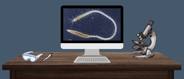
A recent research effort from the University of Tokyo (Japan), led by Toru Miura, has uncovered the mechanics behind one of nature’s strangest reproductive methods: stolonization. The study reveals new information about the developmental mechanics driving this process in Japanese green syllid worms (Megasyllis nipponica), expanding our understanding of developmental biology.
Stolonization describes the process by which an organism develops gonads in the posterior segment of its body, before detaching that segment from the body. Complete with a head, eyes and swimming bristles, this structure is referred to as a stolon. Having detached, the syllid worm’s stolon swims off to find a stolon of the opposite sex, after which it spawns, releasing its gametes before perishing.
It is theorized that this approach provides the organism with a greater chance of encountering sexual partners and successfully reproducing while protecting the ‘stock’ of the organism from potential dangers that may be encountered on the hunt for a mate. However, how these organisms form a new head and eyes in the middle of their bodies is not currently well understood.
 Dinosaur feathers have similar protein composition to bird feathers
In a study on dinosaur feathers, researchers find that fossilization can alter trace protein composition and structure and find similarities to bird feathers.
To address this gap in our understanding, Miura and his team observed the developmental process of stolons in syllid worms. These observations were conducted morphologically, using a stereoscopic microscope and scanning electron microscopy, and histologically, using immunological stains for acetylated α-tubulin. This enabled the team to visualize cytoskeletal microtubules using confocal laser scanning microscopy for the observation of intricate nervous structures. Gene expression patterns throughout the stock and stolon components of the worms were also investigated using RNA extraction and real-time qRT-PCR.
These morphological and histological observations revealed that at the beginning of stolonization, gonads containing the worm’s gametes matured in the posterior end of the forming stolon. This is followed by the formation of the head in the stolon’s anterior segment. Histological investigations revealed that the stolons lacked defined structures present in the stock, such as a pharynx and a proventricle with well-developed muscular tissues; however, a simple digestive tube was present. Neuronal somata with nuclei were clustered in the stolon’s head, forming a basic brain-like structure just before the stolon detached. The stolons also developed functional eyes, antennae and swimming bristles during their development.
The gene expression investigations provided some surprising results for the team. “Interestingly, the expressions of Hox genes that determine body-part identity were constant during the process,” commented Miura. These findings ran contrary to the team’s previous assumption that the Hox genes would vary in expression along the anterior–posterior axis, with an upregulation in anterior Hox genes in the segments of the stolon that form the head.
While surprising, these results do explain why the stolon lacks so many basic physiological features. Instead, only genes for gonadal development were upregulated in the posterior segments before head-determination genes in the anterior segment of the developing stolon were upregulated.
To further elucidate this intriguing mechanism, the team would like to conduct additional studies. “We would like to clarify the sex determination mechanism and the endocrine regulations underlying the reproductive cycles in syllids,” concluded Miura.