I spy with my three-dimensional eye… glaucoma
A novel low-cost 3D imaging device could signal a new advancement in eye screening.
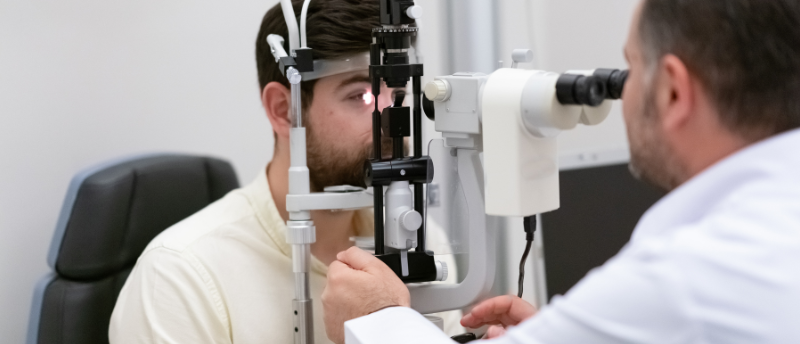
If you want to take a 3D image of someone’s eye, then you had better have quick access to a banker who will do loans on the fly. 3D images are useful for diagnosing retinopathy and glaucoma; however, current instruments (such as optical coherence tomography technology, which uses light to create an image of the back of an eye) can cost up to £100,000 and requires a highly trained specialist. In high-population areas and low-income countries, this can often prove to be prohibitive.
Around 7.7 million people are affected by glaucoma across the globe, which ranks third on the list of common causes of visual deficiency and can result in irreversible blindness. Primary open-angle glaucoma is the predominant form of glaucoma and is predicted to rise from an estimated 52.68 million cases in 2020 to 79.76 million in 2040 in demographics from 40 to 80 years of age†. This represents an escalating burden.
It is good news, then, that researchers at the University of Strathclyde (Glasgow, Scotland) have developed a means of providing 3D imaging using the humble slit lamp (that microscope with the bright light and the chin rest you’ve probably sat in front of at an eye check-up), which is a common piece of technology accessible to optometrists across the globe. The proposed technology is a simple add-on to this standard piece of equipment, providing 3D imaging to any optician. In fact, the technology is said to be simple enough to use in unassisted settings such as pharmacies and, according to researcher Mario Giardini, can take a 3D image in less than a second.
 Reference map of retinal pigment epithelium cells created with AI
Researchers identify five distinct cell subpopulations in the retinal pigment epithelium, which could explain different severities of retinal degenerative diseases.
“The technology has the potential to revolutionize the screening and follow-up within the community of conditions such as glaucoma, as any optometrist, anywhere in the world, could afford it. This work makes eye diagnostics more accessible, reducing inequalities,” Giardini elaborated.
A great deal of ophthalmology depends on 3D visualization, so this new technology represents an advance that is easily implemented to slit lamps and allows for precise measurements to be taken during routine examinations.
This add-on may also be used to provide 3D images of the front of the eye, which is especially valuable for cornea transplants due to the inability of most machines to measure the edge of the cornea. In addition, researchers hope that the technology may eventually be used to detect and measure solid tumors in the eye.
With the initial prototype funded, the team is looking to make the technology available to the medical community and has partnered with the IDCP group (Almere, the Netherlands) to turn it into a medical product.