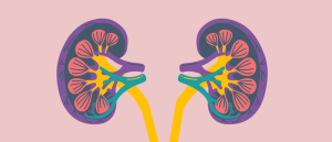
Navigating the nephron with novel hyper-characterized organoids

“You’re on a chemotherapeutic and your oncologist may say that your dosage is X, while a nephrologist is telling you that the proximal tubules in your kidneys can’t handle it. You’re harming them.” This is a reality that many patients may face and a scenario that has driven Nils Lindstrom, leader of a lab in the Department of Stem Cell Biology and Regenerative Medicine at the University of Southern California (USC; CA, USA), on a decade-long odyssey to better understand the nephron (the functional unit of the kidney), the cells that construct it and how we can model it.
Now, with the release of two papers, each first authored by previous PhD students from his lab – MaryAnne Achieng, now working in the Department of Nephrology at the University of California, San Francisco (CA, USA), and Jack Schnell, who now works for Ambry Genetics (CA, USA) – he may have produced a model that can bring the details needed to resolve this fatal catch-22 into focus.
We spoke to Nils, MaryAnne and Jack to learn more about the challenges of investigating the conditions that impact the nephron and their combined efforts to develop organoid models that could change the way we approach these studies.
The nefarious nature of the nephron
“It’s because the kidney is so horrendously complex,” Nils chuckles, almost exasperated in response to the question of why there are so few treatments to address chronic kidney injury or the issues associated with chemotherapy. To really explain this point, Nils details its complex structure, in particular highlighting the role of perhaps the most clinically relevant cell population in the nephron: the proximal tubule.
“At one end it connects to the vasculature,” Nils says of the nephron, “then there’s a long, elegant tube which controls solute homeostasis so that at the other end of the nephron it connects to a collecting duct, which drains the urine.” Right after the filtering structures of the renal corpuscle and the glomerulus, at the end connecting to the vasculature, sits the proximal convoluted tubule. The cells of this structure perform roughly 65% of the reuptake of key nutrients – glucose, salts, amino acids, etc. – from the solution inside the nephron into the body, while the rest is either excreted into the urine, or recovered by other nephron cells.
“To do all this work, they are packed full of mitochondria and need to be incredibly metabolically active to achieve such a high level of reuptake. It also means they are sensitive; you can imagine them running at very high revs to reabsorb so much of what passes them, and as part of this they occasionally reabsorb toxins that are harmful to them.” Among these potentially harmful molecules are chemotherapeutics like cisplatin.
But this is only half the story. For people without cancer, but who have other conditions like hypertension, diabetes or an iron deficiency, the proximal tubule cells are under constant stress, running in a state that, when provoked, can slip into the promotion of an injury response. Unfortunately, the repair mechanism that causes cells to pause, replicate and replace themselves is error prone. “You sometimes end up with a little cell that just can’t escape its repair loop, and instead drives fibrosis, remodeling the regions around it and leading to a loss of renal function.”
Understanding human kidney development
Much of the modeling used at the beginning of the century occurred in mice, a fine model for investigating the basics of kidney development and damage but too physiologically distinct to translate into a detailed understanding of these processes in humans and, ultimately, contribute to clinical advances.
In the early 2010s, Nils, plugged into the field of kidney modeling, knew that new human models were on the way. However, instead of turning straight to IPSCs and trying to create his own models, Nils took the opportunity to join Andrew McMahon in his lab at the University of Southern California to investigate the development of human kidneys.
During this time, Nils operated at the forefront of single-cell transcriptomics and imaging. As human development occurs much slower compared to mice, it allowed Nils and his team to deploy these techniques to interrogate kidney development in real detail.
This work resulted in new maps of human kidney formation that began to explain how the cells of the nephron differentiate depending on their location. “It’s a really elegant mechanism,” Nils explains. “Each progenitor stem cell is recruited from the same stem cell niche, forming a stream of cells moving towards the beginning of the nephron. The first arrivals are subjected to a specific set of receptor ligand interactions that shape their development. This set of interactions is location specific, changing as the cells stack up and compose different sections of the nephron, directing them to form a distal cell or a proximal cell and everything in between.”
With this understanding of human nephron development, Nils was ready to set out to begin the development of kidney organoid models. But first, he would need to secure the right partners to work with.
 SOX9 switch: the key to successful kidney regeneration?
Researchers have uncovered how a SOX9 switch determines the successful regeneration of kidney cells.
Directing differentiation
While Nils was working to provide a detailed blueprint of human kidney development, the first human kidney organoids were released in 2015. However, these models relied almost entirely on the cells self-organizing, a practice that followed on from other early organoid models, such as gut organoids, which self-organize beautifully, but that led to the formation of kidney models with limited morphological relevance.
This presented Nils with a challenge to resolve, and soon, he’d have the tools and team with which to do it. “When I started my lab in 2020, I had the good fortune of MaryAnne and Jack joining as PhD students from the outset. Together we set out to see if we could replicate the rules we found in this blueprint of human development in an organoid model.”
With human kidney organoids already released and in circulation, there was less pressure on the lab to deliver a quick result, and they could instead focus on the finer details of development to produce accurate models. As PhD students, spared the pressures to publish rapidly, Jack and MaryAnne had the time to work meticulously. They were able to conduct a wide range of experiments and utilize innumerable techniques and approaches to create a detailed model that they know inside out.
To do this, they realized they would need to identify which of the receptor–ligand interactions that occur in the different locations of the developing nephron were essential to directing the differentiation of the progenitor stem cells, and which were just noise.
This is where MaryAnne stepped in, reanalyzing countless single-cell RNA sequencing datasets from developing human kidney cells to benchmark the process of nephrogenesis, recognizing the genetic signatures at different stages of differentiation. “The RNA sequencing analysis enabled us,” MaryAnne explains, “to understand more of the transcriptional regulation that happens at different stages of nephrogenesis and to identify what was being underdeveloped in our kidney organoids.”
She also used immunohistochemistry and confocal imaging of human kidneys to get the much-needed context of nephron self-organization, their segmentation, the distinct boundaries between domains and the markers expressed within these boundaries that separated the adjacent cells with completely different cell fates. Immunofluorescent stains were used to establish and demarcate the different segments of the developing nephron. Paraffin sectioning was used to identify post-translational modifications and pinpoint the signaling pathways active in different segments of the human kidney. MaryAnne explained the importance of getting this step right.
“I had to think carefully about how to process my organoids to optimally represent their expression patterns. First, I was learning how to paraffin, embed and section my organoids, which are about 100 microns thick – they’re pretty small – which makes it a little difficult. What helped the most was looking up protocols that already executed the technique I was looking to use in any kind of organoid, adapting them to a protocol that was relevant to my kidney organoids.” MaryAnne has kindly shared her protocol, which you can find here.
With these key pieces of information, the team began learning how to direct the differentiation of progenitor cells into specific cells of the nephron. As these cells developed, they were able to directly compare the cell-fate signatures of the nephron-like cells in their kidney organoids to those documented in developing human kidney cells. “This, combined with the structural data from the imaging studies meant we could establish the benchmark for forming an optimal kidney organoid,” MaryAnne explained.
As the team were now able to define what induced progenitor cell gene signatures should look like at different stages of development and at different locations in the nephron, they were able to optimize nephron induction in the kidney organoids, focusing initially on the cells of the distal portion. They initially used quantitative PCR to monitor signatures as the organoids developed; however, they shifted to bulk RNA sequencing to get a more comprehensive list of the genes activated at different time points.
Using this approach, they were able to successfully produce kidney organoids containing nephrons with a distal identity, composed of cells that matured into thick ascending loop of Henle cells, which compose part of the distal nephron. They then compared the signatures from these cells with published single-cell and single-nucleus RNA sequencing data of both developing and adult human kidneys, confirming that they expressed the genes that encode the ion transporters specific to human distal nephron cells.
 Bat organoid platform reveals more about zoonotic viruses
Engineered mini organs from the most common bat species have been developed to study infections by key viruses, including COVID-19 and influenza.
Producing the proximal tubule…
Working at the other end of the nephron, with the aim of creating a proximal tubular organoid, Jack utilized the information learned to trial a series of different strategies that used small molecule inhibitors – selected for their simplicity and reproducibility – to push cells toward a proximal fate.
The most effective approach, Jack explains “was a short transient inhibition of PI3K kinase signaling at the exact same stage that the organoid nephrons were epithelializing. That intervention boosted Notch activity and pushed cells toward this pre-proximal trajectory. Once we withdrew the inhibitor, the organoid nephrons continued naturally toward an HNF4A positive proximal tubule precursor.”
Using these precursor cells, the team generated proximal-biased organoids, enriched for proximal cells, with nephrons that expressed mature proximal tubule marker genes, carried the right solute carriers and showed molecular uptake. The team could study the development of these proximal-biased organoids using single-nucleus RNA sequencing with high-resolution imaging.
To demonstrate the utility of these models, the team investigated their response to injury, addressing a common approach in similar investigations of toxicity. “Most studies,” Jack explains, “essentially treat organoids with drugs and then point to widespread cell death as an injury response. But of course, any cell will die if you stress them hard enough. And that’s not what happens in patients when they take nephrotoxic chemotherapeutics. Instead, their cellular response to injury is much more sensitive and mosaic.”
So, the team observed how specific pathways, markers and cell states unfolded in real time in response to a toxin, instead of using the ‘blunter’ readout of cell death. Excitingly, they found that the organoids responded to nephrotoxic injury in a similar way to in vivo proximal tubules, replicating the more nuanced response of a real-life scenario.
“I think that seeing all these responses is a major leap forward for the field because it shows that the organoids are no longer just developmental models, they can now serve as functional human platforms for studying proximal tubular disease and testing toxicity,” Jack explained.
And how best to use it…
The main thing about the proximal tubule model that the team wanted to emphasize was just how well characterized it is. “We effectively have a small pharmacokinetic toolkit that is ideal for understanding nephrotoxicity,” Nils underlined. “I’m very biased in my excitement for this, but I really think that studies performing preclinical trials should use this.”
“Another advantage,” Jack highlighted, “is that the proximal biasing intervention happens at the 3D stage, when the organoids are already committed and epithelializing. So that means that you’re working with structured nephrons, with the cells primed to respond in physiologically relevant ways. From a technical standpoint, it makes the protocol quite forgiving because you can reliably stage organoids based on their morphology, then hit them in the right window and get consistent results across different stem-cell lines.”
While it is important to remember that these organoids, including the proximal-biased model, are not meant to be a full artificial kidney, they do provide a consistent human-specific way to study the most disease-relevant cell type of the nephron. What’s more, MaryAnne’s paper provides a framework for researchers to optimize their kidney organoids for the study of different segments of the nephron, with the hope that one day they will be able to construct a full artificial kidney organoid.
So, what is next for Nils’ lab and this model? “In the immediate future, we can now use this model to tease apart how injury repair in the nephron goes wrong, with the ultimate long-term goal of using this as a discovery tool to identify druggable targets and approaches to modulate therapeutics to reduce their nephrotoxicity, because this is simply not a solved problem. There’s not a single good therapeutic out there to help patients with kidney injury, and if my lab is going to have a good translational impact within the next 10 to 15 years, this is where it will be.”
Dr. Schnell and Dr. Lindstrom have reported their disclosures of relevance to the topics discussed in this feature. They are as follows:
- Both Dr. Schnell and Dr. Lindstrom have filed for intellectual property on the proximal-biased system