Imaging dementia: can MRI predict cognitive decline?

New research suggests current neuroimaging techniques could be the future for predicting the development of dementia.7097
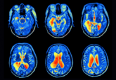
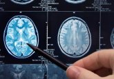
New research from Washington University in St. Louis(MO, USA) and UC San Francisco (CA, USA) suggests that current imaging techniques could one day be used to predict whether an individual will develop dementia. The recent study demonstrated that magnetic resonance imaging (MRI) brain scans could determine if someone would experience cognitive decline in the next 3 years.
Alzheimer’s disease is a form of dementia that affects 5.5 million Americans and currently there is no cure. It is an irreversible disorder, marked by pathological factors that result in neuronal degeneration and cognitive decline, particularly affecting memory and thinking skills.
As of yet there is no way to accurately predict whether an individual will develop Alzheimer’s. “It’s hard to say whether an older person with normal cognition or mild cognitive impairment is likely to develop dementia,” commented lead author Cyrus A. Raji. “We showed that a single MRI scan can predict dementia on average 2.6 years before memory loss is clinically detectable, which could help doctors advise and care for their patients”.
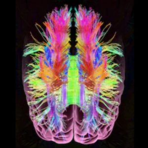
(MRI) brain scans are widely used for structural imaging of the brain and can be vital for diagnosis of many neurological disorders. One technique for MRI scans is called diffusion tensor imaging (DTI), which allows for the analysis of the brains white matter.
The white matter of the central nervous system contains the axons of neurons, the long section of the cell that allows for extended neural communication. The white appearance comes from the myelin sheath, a form of insulation that protects the cell and facilitates the rapid conduction of neural signals. Loss of this myelin slows communication between cells and plays a large role in cognitive decline.
DTI allows for the measurement of the movement of water molecules along the axons and their surrounding myelin. “If water molecules are not moving normally it suggests underlying damage to white tracts that can underlie problems with cognition,” commented Raji.
Researchers studied brain scans taken 2 years previously in groups of individuals who had experienced cognitive decline and those whose cognition had remained the same. They found that those who went on to develop Alzheimer’s showed significantly more damage to their white matter than those who did not.
When looking at the whole brain it was possible to predict cognitive decline with 89% accuracy; when focusing on regions associated with Alzheimer’s it rises to 95%. This is a significant improvement on current predictions that rely on the analysis of risk factors such as the ApoE gene which only has 70-80% accuracy.
“We could tell that the individuals who went on to develop dementia have these differences on diffusion MRI, compared with scans of cognitively normal people whose memory and thinking skills remained intact,” Raji explained.
“What we need now, before we can bring it into the clinic, is to get more control subjects and develop computerized tools that can more reliably compare individual patients’ scans to a baseline normal standard. With that, doctors might soon be able to tell people whether they are likely to have Alzheimer’s develop in the next few years,” he added.
Though there are currently no treatments that can prevent or delay Alzheimer’s development, these results can be highly beneficial for those at risk as they are given the ability to be more prepared for their future.


