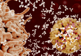
Coronavirus’ covert defense mechanisms discovered by helium ion microscopy
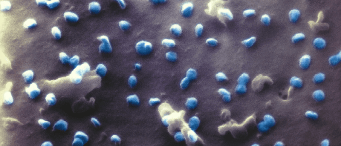
Visualizing the SARS-CoV-2 virus has been integral to discovering ways of combating the disease in the past year; using a helium ion microscope means this can be done with even more clarity. A team of scientists from the Faculty of Physics at Bielefeld University (Germany) have successfully used a helium ion microscope to image the SARS-CoV-2 virus. This method of imaging produces much clearer images than electron microscopy due to the lack of a thin metal coating (necessary in electron microscopy), which can interfere with visualizing the reactions between the virus and the host cell. “The study shows that the helium ion microscope...
To view this content, please register now for access
Join our member community for FREE to access a collection of journal and online-only features, including:
- Exclusive access to educational videos, eBooks and insights into top BioTechniques journal articles
- The latest news and journal updates delivered straight to your inbox when you want it
- Personalized recommendations for the latest member-exclusive podcasts, interviews and expert opinions
- Priority registration to webinars, panel discussions and events
- Access to competitions and journal publication discounts, including 10% off open access fees when you sign up today!