New ultrasound method ensures dolphin pregnancies are going swimmingly
A novel technique allows researchers to ultrasound dolphins in the same way as humans, which may contribute to increased reproductive success.
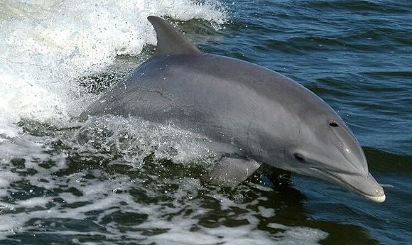
The 2010 Deepwater Horizon oil spill resulted in large-scale oil contamination of the northern Gulf of Mexico and consequentially caused long-term health effects and reduced reproductive success in the population of bottlenose dolphins (Tursips truncatas) in the area. Since then, there has been a huge effort from conservationists to increase reproductive success within the species.
Now, a team from the National Marine Mammal Foundation (CA, USA) has developed a novel ultrasound technique for assessing dolphin fetuses at all stages of gestation, which mirrors the exam used in humans. This breakthrough in dolphin medicine may help to improve reproductive success and elevate the species’ conservation status.
“We can now re-create the human 20-week fetal ultrasound exam in dolphins, which means we can better understand the health challenges dolphin mothers and their babies are facing,” explained Cynthia Smith, lead author on this paper. “This is a game-changer for the conservation of bottlenose dolphins and other small cetaceans around the world.”
The technique was developed through a longitudinal cohort study, published recently in Veterinary Radiology and Ultrasound, in which the researchers closely followed 16 healthy pregnancies of 12 dolphins in human care over a 7-year period (2010-2017).
In total, 203 ultrasound examinations were included in the study and 70 ultrasonographic parameters were assessed for each image. The researchers identified several metrics that could accurately predict fetal age, including fetal biparietal diameter, aortic diameter, blubber thickness and thoracic width and height.
- Natural selection in utero
- New insights into sexual differentiation in frogs
- An ultrasound peek into live cells
From this, the researchers determined the ultrasonographic characteristics of a normal pregnancy at each trimester, which allows for easier identification of abnormalities in pregnancy. The team also provided an accurate equation to calculate the predicted due date in Tursiops which should be useful in advising the timing of clinical decisions as parturition approaches.
“This new technique can be performed in a matter of minutes and provides a wealth of information about the health of the dolphin fetus,” remarked co-author Marina Ivančić. “We are thrilled to make this technique widely available to veterinarians and radiologists, which has the potential to elevate dolphin medicine globally.”
Future applications of this method will allow veterinarians to identify in utero abnormalities and maternal illness, as well as help to improve our understanding of reproductive failure in both wild and managed dolphin populations. Overall, this should assist conservationists’ efforts to regain stability in the populations affected by the oil spill.
“This advanced ultrasound technique is allowing us to diagnose problems as early as the first trimester of pregnancy in dolphins,” commented study author Forrest Gomez. “That gives us a chance to determine if there is something that could be done to save the pregnancy, which could prove critical for populations of dolphins and porpoises that are at risk.”