Tips and best practices to optimize your exosome labeling
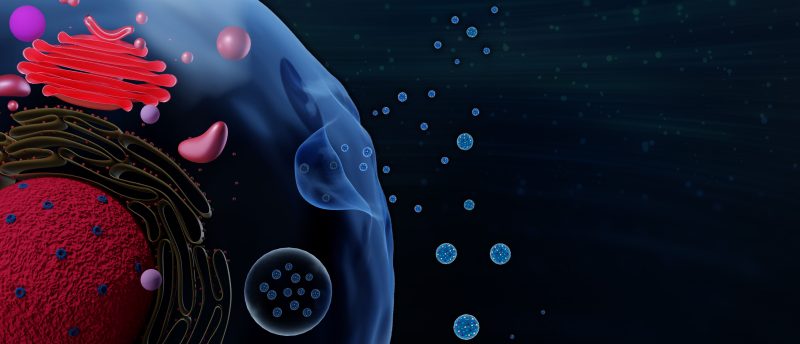
Extracellular vesicles (EVs) are small, membrane-bound particles secreted from cells and thought to function as cellular messengers, carrying cargo from one cell to another. In biomedical research, exosomes and their cargo are used as diagnostic biomarkers for cancer and other diseases. However, the isolation and detection of exosomes can be extremely challenging due to their small size (~30-200 nm in diameter, similar to most viruses). New methods and tools are constantly being developed but it can be difficult to know which to use.
In this tech tip, Biotium scientists share their expertise for optimal fluorescent staining and detection of exosomes by flow cytometry.
This article reveals:
- A detailed comparison of exosome isolation methods
- Tips for using fluorescent antibodies or membrane dyes to label exosomes
- Best practices for flow cytometry detection of exosomes
And much more!
This content was provided by Biotium.
You might also be interested in...