Molecular imaging identifies C. diff structure
A novel combination of multiple advanced molecular imaging techniques has led to the discovery of two molecular structures, which could provide a therapeutic target for C. diff infections.
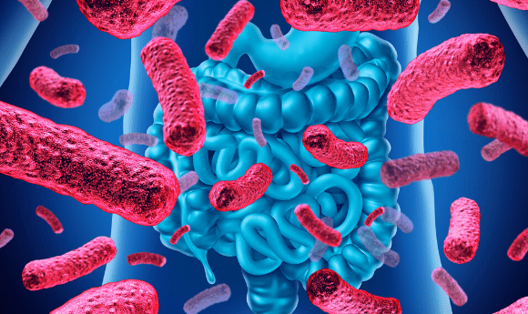
A recent publication in Proceedings of the National Academy of Sciences, has potentially uncovered two targets for drug development against the virulent pathogen Clostridium difficile (C. diff). Although C. diff usually resides in the gut, overuse of antibiotics can disrupt the natural flora allowing C. diff to proliferate, which can cause diarrhea, nausea and internal bleeding. In the US, over 500,000 cases are reported annually, resulting in 15,000 deaths.
“The most dangerous strains of C. diff release a binary toxin that first binds to cells and then creates a pore-forming channel that allows the toxin to get inside and do harm,” explained author Amedee de Georges (City University of New York, NY, USA).
“We were able to combine several increasingly popular biophysical imaging techniques to visualize and characterize every atom of this binary toxin and show us where they are positioned. These details provide a critical and extremely useful starting point for designing drugs that can prevent C. diff infection.”
In an attempt to elucidate this toxin, researchers utilized a variety of structural and biophysical approaches, including cryogenic electron microscopy, X-ray crystallography, nuclear magnetic resonance and small angle X-ray scattering.
- Could insulin prevent the spread of dengue, Zika and West Nile Virus?
- Have researchers found a treatment for severe dengue disease?
- Exploring the future of fecal microbiota transplantations
Initially, the researchers believed that the C. diff toxin was similar to the anthrax toxin – consisting of two components, an enzymatic subunit combined with a pore-forming subunit. However, cryogenic electron microscopy enabled the researchers to visualize the pore-forming subunit in two different conformations.
“We observed two similar but distinct forms of the C. diff toxin — one where we see the pore-forming channel and one where it is invisible” commented author Xingjian Xu (City University of New York). “This gives us clues as to how to prevent the formation of the channel and stop the bacteria from entering the cell,” Xu continued.
Additionally, the team identified a novel calcium-binding structure, which may be critical in C. diff toxin stability. In the hope of curing this infection, the novel identification of these two structural components of the C. diff toxin may provide the basis for future drug development.