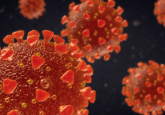
New single-molecule imaging technology could open a window into SARS-CoV-2
Obtaining clear, informative imaging data for SARS-CoV-2 could prove vital in unlocking the virus’s Achilles’ heel, but so far obtaining this information requires bulky, expensive equipment that can prove inoperable in BSL3 or 4 Labs, meaning studies are carried out on “virus-like” particles. Could a new imaging technology, allowing virologists to conduct single-molecule imaging in a BSL3 or 4 lab, help unlock new secrets about SARS-CoV-2?
If you would like to keep up to date with our content on coronavirus, you can sign up for our site here, where you can subscribe to our newsletters for free!
Please introduce yourself and your institution.

Ricardo Bastos
My name is Ricardo Bastos and I am the Head of Applications at ONI (Oxford, UK). We’re a start-up biotech company that pioneers the next generation of super-resolution microscopes. Most of us come from academic backgrounds and have first-hand experience working in the lab with a variety of microscopes. We bring that experience into the design process, always keeping the end-user in mind. More than just designing and building microscopes, we engage in constructive research collaborations between academia and industry as well as conducting our own research in-house. Our mission is to make advanced fluorescence microscopy accessible to all scientists.
Please can you tell us about the role of single-molecule imaging techniques in virology?
With single-molecule imaging, we can study individual molecules one at a time. This enables us to dissect every different confirmation, structure, function, and even subcellular location of a molecule. These finer details are lost in low-resolution conventional fluorescence microscopy.
There are several forms of single-molecule imaging that can be deployed to address specific research questions. Some provide primarily spatial information and others provide temporal or dynamic information. Spatial information can be gained with techniques like super-resolution microscopy and single-molecule FRET, whilst dynamic information is obtained from techniques like single-particle tracking.
Both spatial and dynamic information are essential for the study of viruses. Most viruses are less than 300 nm in diameter, which means they appear as blurry spots in conventional microscope images. However, with super-resolution microscopy, the structure of a virus can be resolved. Combined with different fluorescence probes, we can now visualize the virus envelope, capsid, and genomic material. With other live-cell single-molecule techniques the real-time tracking of viruses can give us insight into how viruses infect cells, hijack cellular machinery, and re-assemble into new viruses.
Can you give us an overview of the different techniques employed in single-molecule imaging studies of viruses?
One of the main classes of super-resolution imaging is single-molecule localization microscopy. dSTORM (direct Stochastic Optical Reconstruction Microscopy), or variations like PALM (Photoactivated Localization Microscopy) and DNA-PAINT (DNA points accumulation for imaging in nanoscale topography), enable fluorescently tagged molecules to be imaged with 20 nm resolution. This is achieved by making fluorophores blink so that the exact position of each fluorophore can be pinpointed in space. Once all the fluorophores have been detected, a super-resolved image is formed by combining all the precise localizations of each fluorophore.
Alternatively, if you want to study the conformation of proteins or the interactions of neighboring molecules between 1-10 nm, single-molecule FRET (Förster resonance energy transfer) can be used as a molecule ruler. Finally, single-particle tracking enables the dynamic, real-time localization of viruses during assembly and maturation, envelopment, and release.
Ultimately, single-molecule imaging can help resolve the underlying mechanism of viral infection. These information-rich techniques allow us to probe the dynamics, stoichiometry, and conformation of viruses and their components.
What are some of the challenges of implementing these techniques?
In the field of viruses, researchers face inherent health risks, requiring that work be conducted in secure and restricted facilities. This greatly restricts the instrumentation and techniques that virologists can use, as most microscopes are too large to be placed in a biosafety level (BSL) 3 or 4 containment facilities. Therefore, many researchers are forced to study virus-like particles as opposed to actual viruses. This raises the concern of whether the conclusions extrapolated from research on virus-like particles are transferable to the virus itself.
In addition, one of the main barriers to single-molecule imaging is that these techniques are mostly used by specialists who’ve trained on and worked with advanced instruments for many years. Single-molecule techniques require complex instrumentation, dedicated infrastructure including a dark room and optical table, and often complicated experimental design. Many single-molecule microscopes are optimized for specific techniques, which then requires researchers to have access to and training on many different instruments. Unfortunately, these challenges make single-molecule imaging inaccessible to many researchers.
 Single-molecule imaging with Bruno da Rocha-Azevedo
At ASCB 2019 we discussed the single-molecule imaging of the receptor VEGFR-2 with Bruno da Rocha-Azevedo. Bruno took us through the technique and discusses its challenges, such as the difficulties of imaging 3D tissues and samples, and his predictions for what the future holds for single-molecule imaging and analysis of 3D samples.
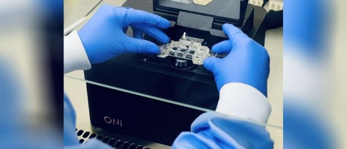
What is the Nanoimager device and how does it begin to address these challenges?
We wanted to build a microscope that would fit the workflow of scientists, instead of relegating them to dark basement rooms. The Nanoimager is essentially a small black box with a footprint the size of an iPad. Its compact and unique design allows researchers to image pretty much anywhere, directly on the lab bench with the lights on and without the need for bulky and expensive optical tables. This includes BSL3 labs which is unheard of for standard super-resolution microscopes. Now, virologists are no longer limited to studying virus-like particles and can focus on understanding the viruses themselves.
We also equipped the Nanoimager with the capabilities to perform a number of different techniques, including dSTORM, single-particle tracking, and smFRET that I mentioned earlier. This gives users the flexibility to implement a wide range of techniques without having to be trained on multiple instruments.
Finally, a key aspect of accessibility is price. Quite frankly, the Nanoimager is about half the price of other microscopes of this caliber, which enables more labs around the world to take advantage of single-molecule imaging for their research.
What information are you able to obtain with this device that you wouldn’t be able to with existing techniques?
The compact size of the Nanoimager means that imaging can be done practically anywhere. During the current COVID-19 crisis, I have even set-up a Nanoimager in my home that I’m using for remote demonstrations. This is a game-changer, as it enables us to conduct super-resolution imaging in the field.
As we’ve recently seen, there is a huge demand for diagnostic virus testing. Typically, the gold standard is validation by EM, which requires the use of large and expensive equipment, on top of complex sample preparation. However, with COVID-19, it is essential that accurate testing can be deployed to workplaces, transit centers, and communities. In the future, we believe that single-molecule imaging will play a much more prominent role in clinical settings as it would enable rapid diagnostics to be performed onsite at locally designated hubs.
Do you have any tips for best practice when conducting single-molecule imaging studies?
This is very dependent on the kind of question you want to answer and the kind of experiment you would like to conduct. For example, with the increased resolution it becomes essential that every single molecule of interest is labeled. If labeling is too low, we won’t be able to obtain an accurate picture of the imaged structures.
In addition, each single-molecule technique requires something different out from the fluorophores to obtain high-quality images. For example, dSTORM imaging requires robust blinking, while live-cell tracking requires stable fluorescence with minimal photobleaching. When designing experiments, it is therefore important to choose fluorophores specifically for each technique based on their unique properties.
Where do you see single-molecule imaging in virology in the next 5 years?
While there has been an increased interest in the use of single-molecule imaging in virus research, I would love to see more virologists, and research labs in general, using this technology in their day to day work.
As a company, ONI is making single-molecule techniques more accessible, both in terms of ease of use and safety standards. The accessibility of single-molecule imaging is governed not only by the hardware but also by its software and analysis tools. To this end, we are creating a platform that enables scientists anywhere in the world to access and analyze their data. Through collaborations with academic and industry partners, we are producing sophisticated and powerful, yet easy to use, tools that scientists can use to solve some of the world’s most pressing problems.