SHANEL: new technique shows promise for 3D-printed human organs
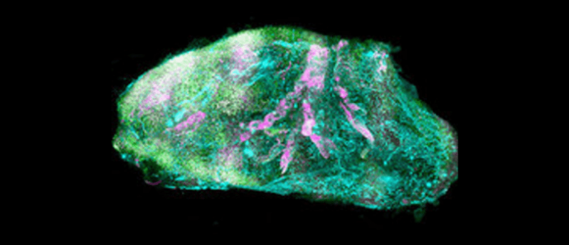
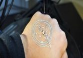
A new microscopic imaging approach has made intact human organs transparent for the first time, raising hope for the development of 3D-printed human organs suitable for transplant.
Advances in tissue clearing approaches have allowed researchers to observe the cellular structure of transparent, 3D mouse organs. Knowledge of this structure is essential for the development of 3D-bioprinted organs suitable for use as models and in organ transplant.
However, human organs are too ‘stiff’ for the mouse approach owing to the build up of insoluble molecules in older tissues. This means that the detergents used in mice do not work in humans.
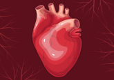
Now, a team from Helmholtz Zentrum München (Germany) has taken a different tack, revealing details of the human eye, thyroid, kidney and transgenic pig pancreas.

Interested in this piece? Click here to check out the rest of our spotlight on 3D cell culture.
This involved the discovery of a detergent called CHAPS, which makes centimeters-deep small holes throughout the organ, through which solutions can be administered to make the organ transparent.
Then, a new laser-scanning microscope was developed in collaboration with Miltenyi Biotec (Bergisch Gladbach, Germany), which can image large organs – the “Ultramicroscope Blaze”.
Finally, the team developed a deep-learning pipeline able to analyze the millions of cells then cleared.
In its entirety, this new technology is called SHANEL – Small-micelle-mediated Human organ Efficient clearing and Labeling.
The team is now using the technique to map some of the major organs of the human body, beginning with the pancreas, heart and kidney.
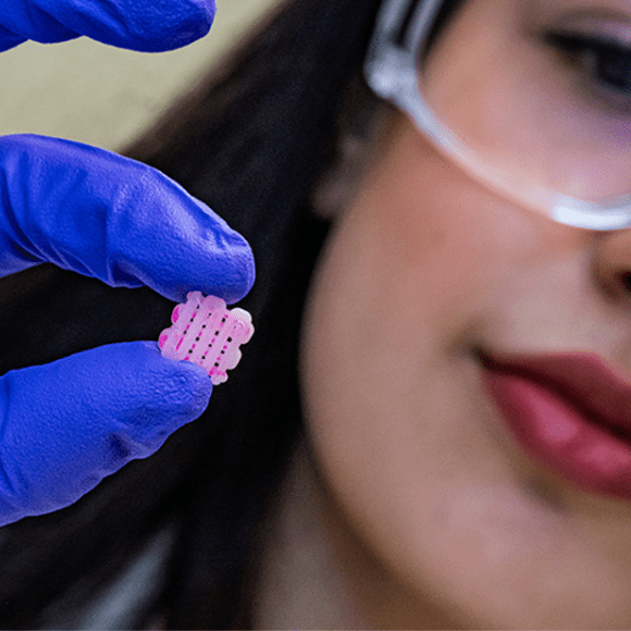 Tissue engineers get into the groove
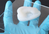
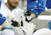
A new method promises to help heal injuries using grooved 3D-printed tissue-engineering scaffolds that can be seeded with living cells. The grooves protect the cells, while also allowing them to be seeded using a bioink in a layered manner alongside bioactive molecules.