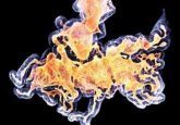
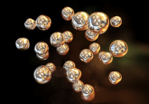
Cryo-EM could be used to visualize proteins at the atomic level

Cryogenic electron microscopy (Cryo-EM), the electron microscopy technique applied to cryogenically cooled sample, has been used much over the past decade as a reliable imaging technique. Now, researchers from two studies undertaken at the Max Planck Institute for Biophysical Chemistry (Göttingen, Germany) and the Medical Research Council Laboratory of Molecular Biology (MRC-LMB) in Cambridge, UK, have made a breakthrough in achieving imaging at the atomic level in Cryo-EM. Currently, visualizing structures such as proteins at such a high resolution so as to better understand their structure in relation to function, is not easily achieved by other standard imaging techniques, such...
To view this content, please register now for access
Join our member community for FREE to access a collection of journal and online-only features, including:
- Exclusive access to educational videos, eBooks and insights into top BioTechniques journal articles
- The latest news and journal updates delivered straight to your inbox when you want it
- Personalized recommendations for the latest member-exclusive podcasts, interviews and expert opinions
- Priority registration to webinars, panel discussions and events
- Access to competitions and journal publication discounts, including 10% off open access fees when you sign up today!