It’s not just carrots that help us see in the dark
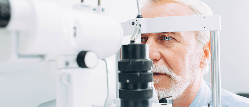
Researchers have characterized the three-dimensional structure of the cyclic nucleotide-gated (CNG) ion channels that help us see in dim light, hoping this will lead the way to new medical treatments.
“It’s thanks to the rod cells in our eye that we can observe the stars in the night sky,” says Jacopo Marino (Paul Scherrer Institut; Villigen, Switzerland), who led this study. “These photo cells are so sensitive to light that they can detect even a single photon reaching from a very remote part of the universe – a truly incredible feat.”
The CNG ion channel is embedded in rod cell membranes, and controls which particles can pass through to the interior of the receptor cells by opening and closing depending on the amount of light.
In dim light, this channel is completely open; however, when light hits the eye, a cascading set of processes is initiated leading to the CNG closing. When this ion channel closes, positively charged particles like calcium ions can no longer enter rod cells. This causes a set of electrochemical signals to travel via nerve cells into the brain’s visual cortex where a visual impression is produced, like a flash of light.
“The idea of solving the structure of this channel dates back to nearly 20 years ago when Gebhard Schertler and Benjamin Kaupp already collaborated on this topic,” explains Marino, Schertler and Kaupp are both co-authors of this new study.
 Cataract surgery could reduce dementia risk: it’s clear to see
Researchers have found that cataract extraction surgery is associated with a 30% lower risk of dementia.
It took 2 years for PhD student Diane Barret to extract enough of the ion channel protein from cows’ eyes to study. Barret explains this is because the protein decomposes very quickly and is only available in small quantities in the source material. “We were both too stubborn to simply give up,” says Marino, “but in the end that stubbornness paid off.”
Once enough of this protein was collected, cryo-electron microscopy was used to visualize the ion channel in its closed native state, unlike in previous studies. The researchers found that the CNG channel is made up of four parts: three subunit As, and one subunit B. They observed that subunit B had a side arm, arginine, sticking out into the channel, acting like an additional gate across the channel and further narrowing it. “No one expected that – it came as a total surprise,” explains Barret.
The researchers noted that this extra barrier from subunit B is found in all animals with the same amino acid at the same position on the protein, which has been maintained during evolution and is therefore thought to be essential for the channel to work correctly.
A better understanding of how we see could help treat certain genetic disorders that lead to blindness, such as retinitis pigmentosa, which is thought to be caused by a genetic defect resulting in an incorrectly formed CNG channel. This occurs as the reactions leading to the channel closing cannot be initiated, which affects the electrochemical balance causing photoreceptor cells to die off and eventually results in blindness.
“If we could find molecules that affect the protein in such a way that the channel would completely close, we could prevent the cells from dying – and thus stop people going blind,” explains Marino. Knowing the structure of this channel means such molecules can now be looked for.
Aside from answering this 20-year-old question on the structure of the CNG channel, it is hoped this new insight into vision will advance the development of treatments for certain genetic disorders.