First cell atlas for navigating kidney diseases

Researchers have shone a light on the kidney immune system by developing the first cell atlas of the human kidney, which will have important implications for treating kidney disease.
Chronic kidney disease is a serious condition affecting more than 850 million people worldwide and can often lead to fatal kidney failure. The immune system plays an important role in determining how kidney tissues respond to damage, but relatively little is known about how this works.
Now, however, researchers have created the first cell atlas of the human kidney immune system. This shows where different immune cells are located in the kidney and gives a much greater insight into what happens when kidney disease or damage occurs.
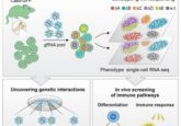
Using single-cell RNA sequencing, the researchers sequenced the activity of 67,471 individual kidney cells from different stages of life. These cells were then mapped to generate the spatiotemporal topology of the human kidney to determine how the immune system develops and is organized.
“We have created the first map showing how the immune system in the kidney develops in early life and how that changes as we mature into adults” commented co-lead author Muzlifah Haniffa, professor of dermatology and immunology at Newcastle University (UK).
The study, published recently in Science, demonstrates how our kidney immune system initially develops during fetal stages and then progresses and strengthens after birth and as we continue to mature into adults.
- Interspecies organ donation
- Miniaturizing models of liver disease
- Gut instinct: how to treat allergies
Macrophages were found to be present in the kidney at the earliest stages of development, but other active immune cells only seemed to develop postnatally. After birth, the kidney acquires transcriptional programs that promote infection defense capabilities.
“We uncovered the very earliest cell types in the developing kidney – these macrophages that live in the kidney throughout life are important for protecting us against infection,” Haniffa explained.
Researchers were also able to identify the pelvic epithelium as the area of the kidney most vulnerable to bacterial infection. This area consequentially had the highest expression of immune genes in the mature kidney, but not the fetal kidney.
The results from this study provide a much deeper understanding of the kidney immune system, and will have important implications for managing, diagnosing and treating kidney disease, including helping clinicians to understand why kidney transplants can be rejected.
However, the researchers feel that the impact of this study could extend even further than just treating kidney disease.
“Mapping the human kidney brings us one step closer to producing the Human Cell Atlas – a Google map of the 37 trillion cells in the human body,” remarked Sarah Teichmann, co-lead author and researcher from the Wellcome Sanger Institute (Cambridgeshire, UK). “We will discover new cell types and uncover how our cells change over time, learn how and why we age and what happens when we get a disease. The Human Cell Atlas will be a free online resource, for anyone to use.”


